
Services · Temara, Morocco
Consultations, examinations and care
From your first visit to follow-up, explore the examinations available at the practice and the care coordinated for your situation. Every examination and treatment requires an individual medical indication.
Services
Your visit, step by step
A clear process, from your arrival to the medical plan.

Welcome & patient record
The secretary welcomes you and enters your administrative information into the practice system.
Appointment & numbered queue
Your appointment for the day is checked and a queue number organises the waiting area. The order may change if a medical emergency arises.
Medical history
In the consultation room, the doctor discusses symptoms, concerns, age, medical history, medication and risk factors, and clarifies whether the concern relates to urology, fertility or sexual health.
Clinical examination
An examination tailored to your concern, including palpation and vital signs when needed, with respect for your privacy.
Tests & explanations
Relevant examinations are proposed according to the findings. The doctor explains the results, treatment options and follow-up. Not every test is necessary for every patient.
At the practice · when indicated
Examinations at the practice
Complementary tests selected according to your symptoms and clinical assessment.
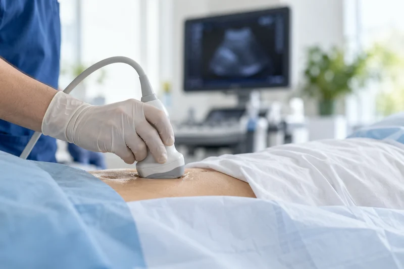
At the practice · when indicatedUrinary ultrasoundKidneys, bladder and prostate, as appropriate.
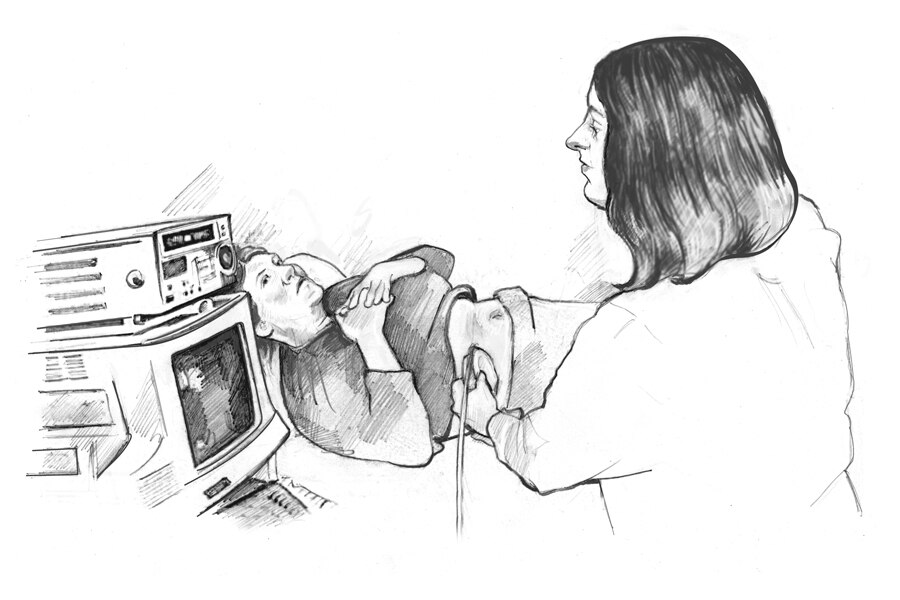
Kidney, bladder and prostate ultrasound for men, and kidney and bladder ultrasound for women, using a latest-generation ultrasound system. Bladder assessment requires sufficient bladder filling.
Post-void residual measurement assesses the urine remaining after urination and complements the examination when indicated.
At the practice · when indicatedUroflowmetryMeasuring how the bladder empties.
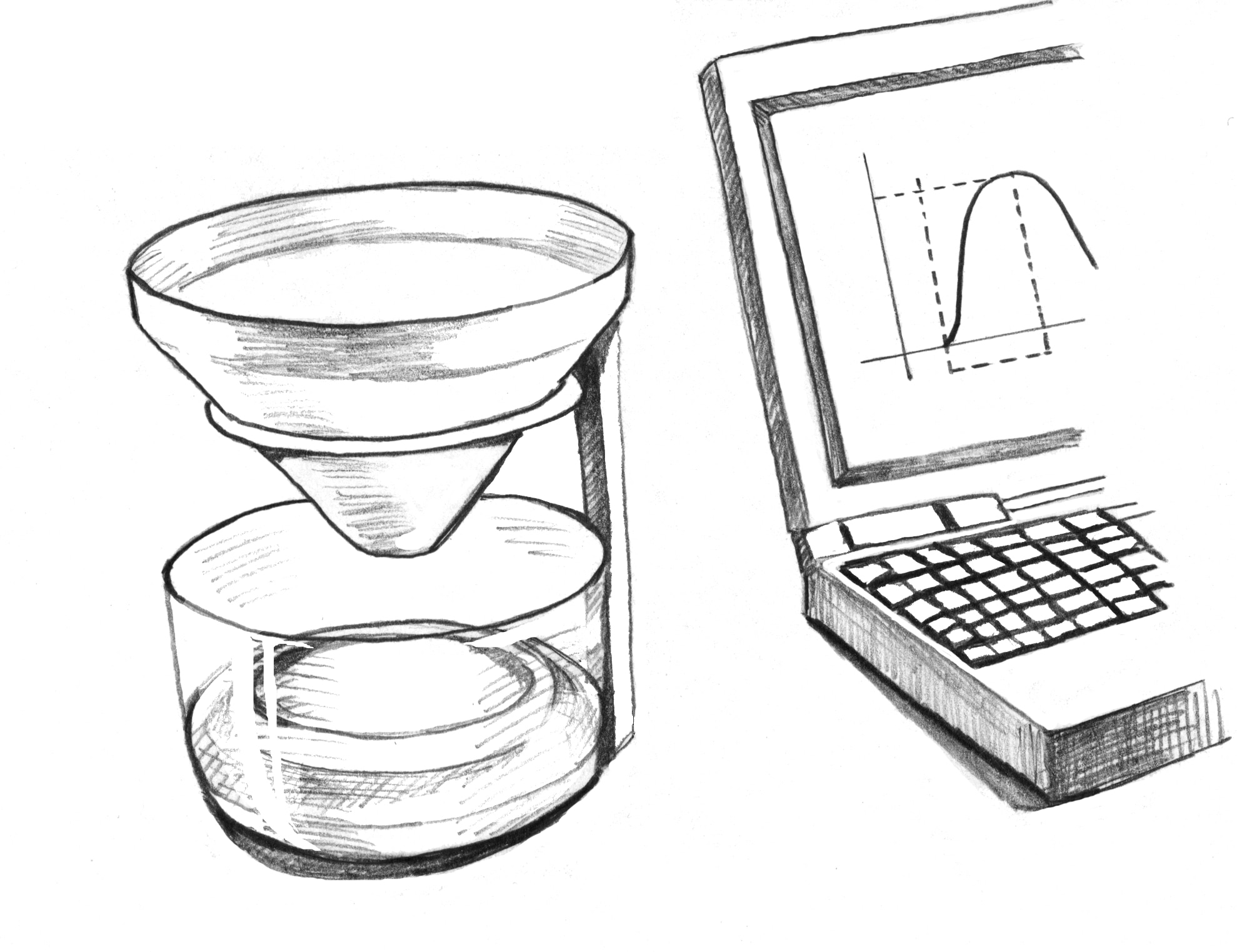
This non-invasive test records urinary flow rate, the flow curve and voided volume. It supports assessment of urinary symptoms in men and women and monitoring of treatment for benign prostate enlargement.
Reduced flow may suggest obstruction, urethral narrowing or impaired bladder contraction. It cannot establish the cause alone: findings are interpreted with symptoms, residual urine and further tests when needed.
Preparation and documents for this test
At the practice · when indicatedFlexible cystoscopyLooking directly inside the urethra and bladder.
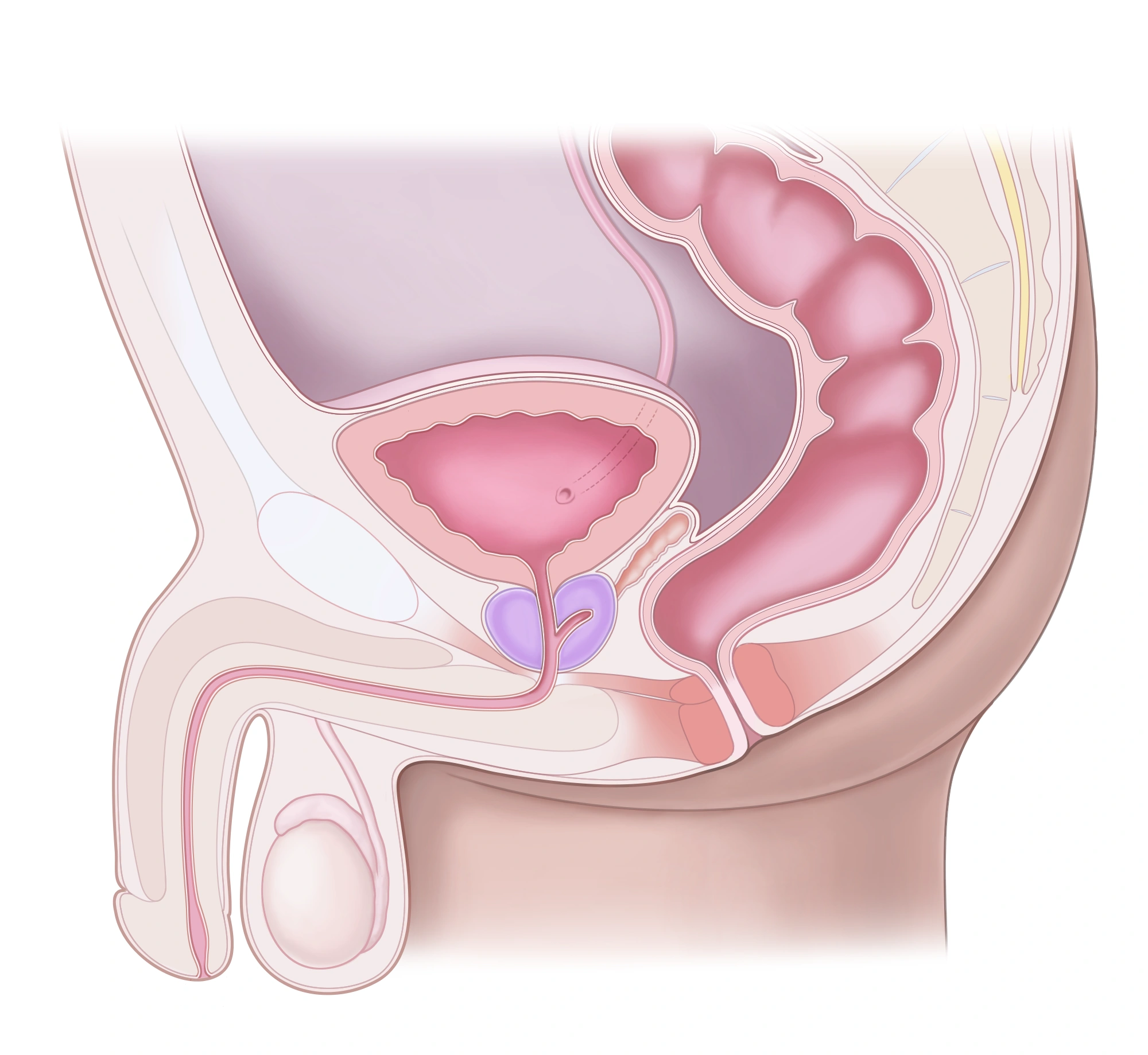
Reading this diagram
The prostate sits below the bladder. The urethra passes through the prostate and then the penis. The rectum lies behind them.
Olympus flexible cystoscopy can be performed at the practice after checking the indication and safety conditions. It helps investigate blood in the urine, urethral abnormalities or suspicious bladder lesions.
It may contribute to assessment of bladder pain syndrome or interstitial cystitis, but is not a routine screening test for these conditions. Biopsy depends on the lesion and is performed in an appropriate setting; it is not required for every patient.
Preparation and documents for this test
At the practice · selected patientsUltrasound-guided prostate biopsyDiagnostic tissue sampling under local anaesthesia.
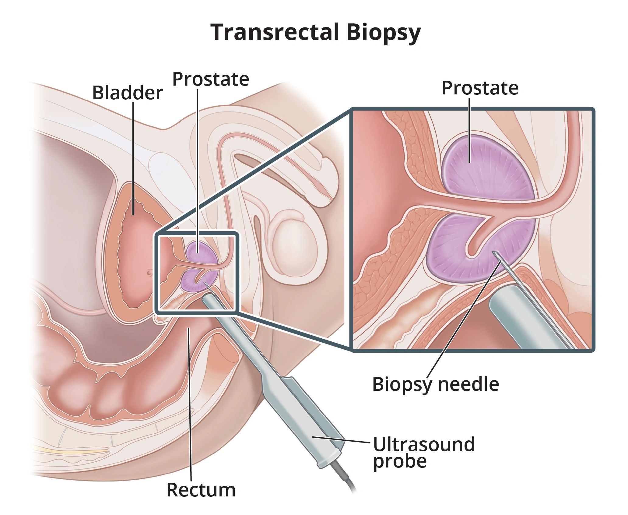
Available at the practice for selected patients following review of general health, medication, infection and bleeding risks. The decision follows prostate assessment; MRI is usually performed before biopsy when localised cancer is suspected.
Ultrasound guides the sampling. The EAU favours the transperineal route to reduce infection risk; the route and targeting method are explained beforehand. Written aftercare instructions and follow-up of pathology results are provided.
Preparation and documents for this test
Transrectal biopsy: preparation, procedure and follow-up · FR / العربية
At the practice · when indicatedScrotal ultrasound with DopplerVaricocele, masses, hydrocele and epididymal cysts.
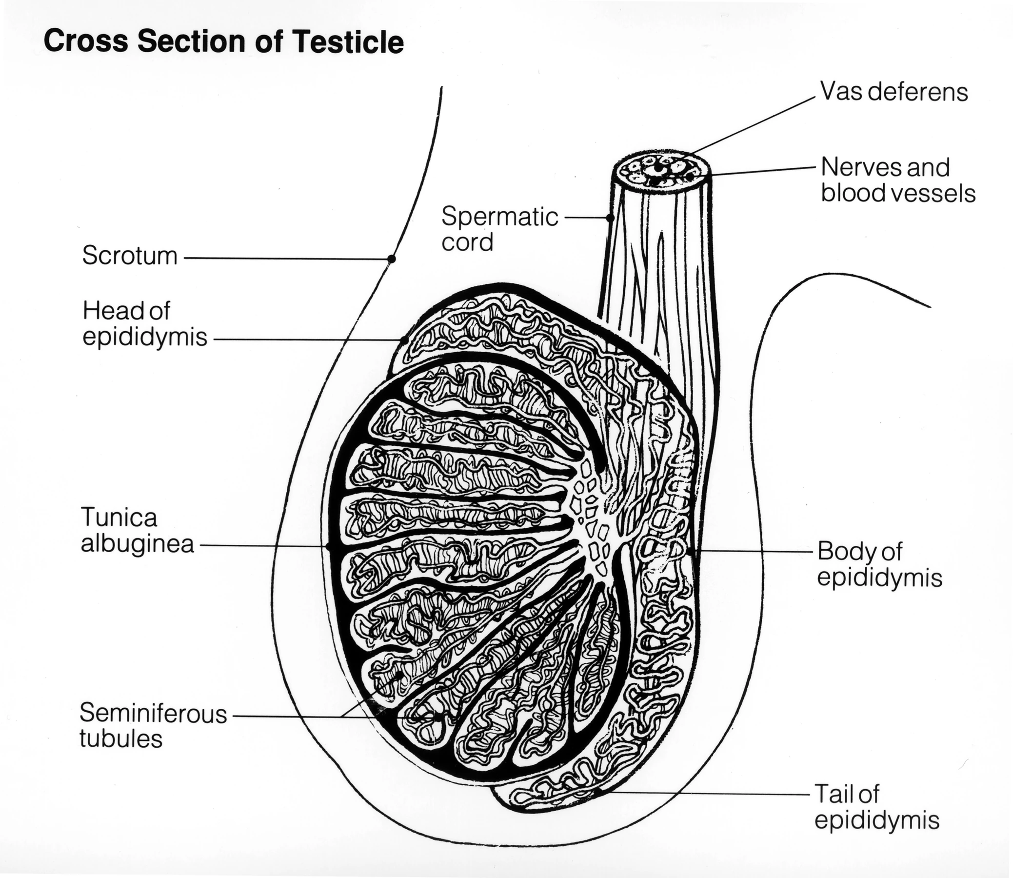
Assessment of the testes, epididymides and scrotal blood flow. It complements examination for pain, swelling, a suspected tumour or varicocele; it is not routine ultrasound screening for every man.
Varicocele grades 1, 2 and 3 are clinical: palpable with straining, palpable at rest, then visible and palpable at rest. Doppler additionally assesses venous reflux and testicular volume.
At the practice · when indicatedRenal Doppler ultrasoundVascular assessment for a specific clinical indication.

Renal Doppler complements ultrasound when assessment of renal blood flow is relevant. Its use depends on the clinical question and other examination findings.
It is distinct from scrotal Doppler used for varicocele assessment. Further imaging or specialist advice may be needed depending on the findings.
THE SPECIALTIES
Care organised by specialty
Consultations, assessment and follow-up are coordinated at the practice. Surgery, radiotherapy and treatments requiring dedicated facilities are arranged in a clinic or specialist institution.
Consultation & assessmentProstate, urethra & urinary symptomsUnderstanding the cause of urinary difficulties.
Reading this diagram
The prostate sits below the bladder. The urethra passes through the prostate and then the penis. The rectum lies behind them.
Assessment of difficulty urinating, a weak stream, frequent urination, incomplete emptying and functional urinary problems in women and men.
Clinical examination, ultrasound and uroflowmetry guide the assessment. Further tests and treatment are tailored to the findings, including prostate enlargement or suspected urethral stricture.
In a surgical clinic · when indicatedProstate resection & endoscopic surgeryRelieving obstruction caused by benign enlargement.
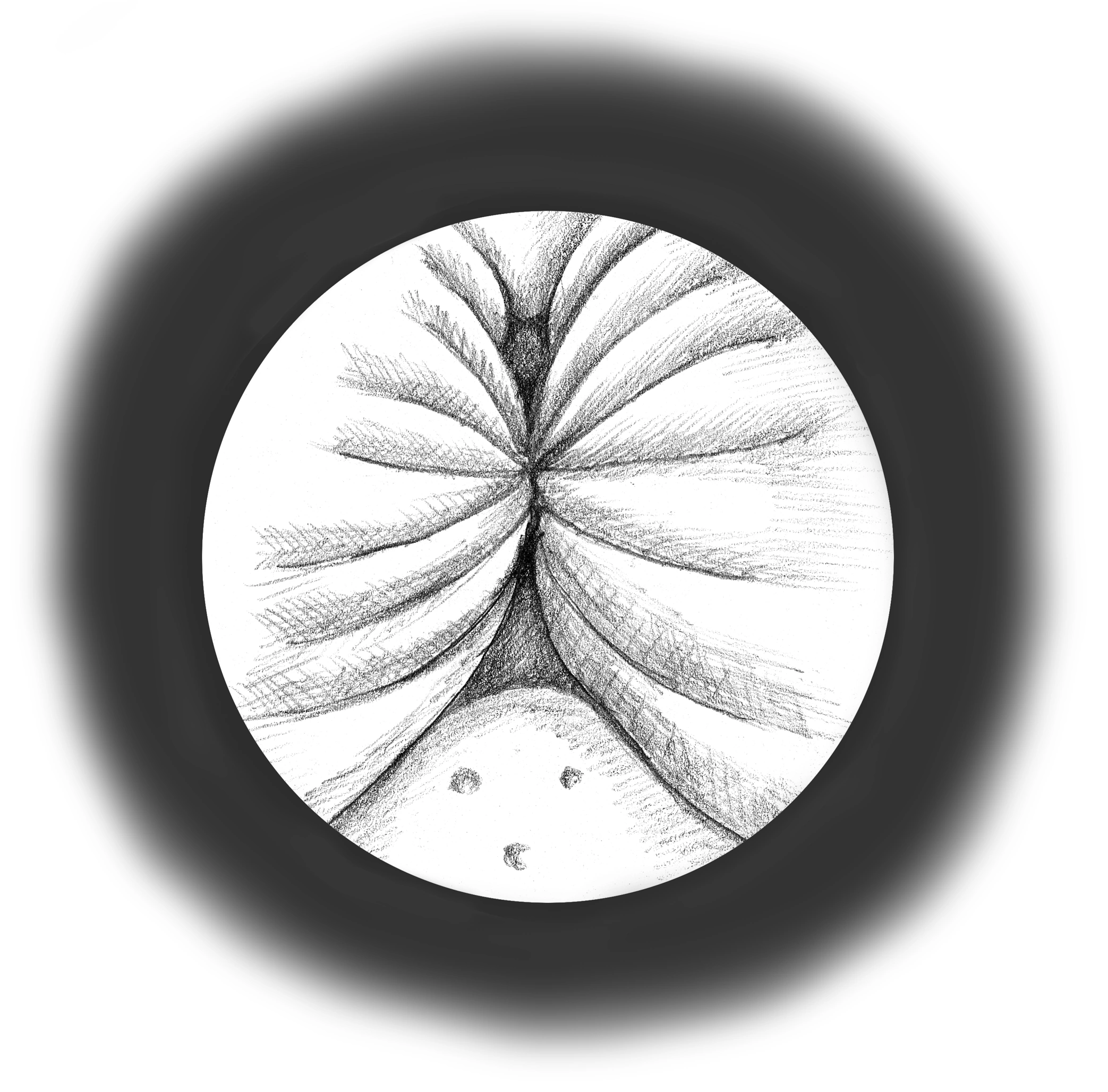
Reading this diagram
This view looks from inside the urethra. The walls are pushed together, leaving a narrower passage for urine.
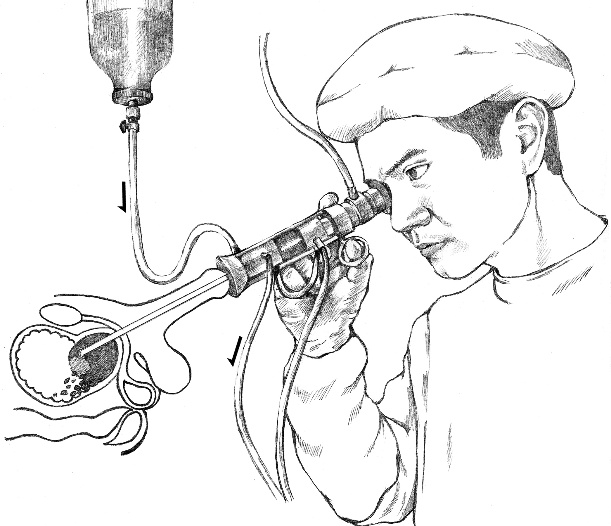
Procedures in a surgical clinic when symptoms or complications of benign prostate enlargement warrant surgery. The approach depends on prostate size, medication, bleeding risk and available facilities.
Transurethral resection, including bipolar resection, and enucleation or laser alternatives are discussed where appropriate. Urinary recovery, possible effects on ejaculation and alternatives are explained. This differs from prostatectomy for cancer.
In a surgical clinic · when indicatedBotulinum toxin for overactive bladderInjections into the bladder wall in a surgical clinic.
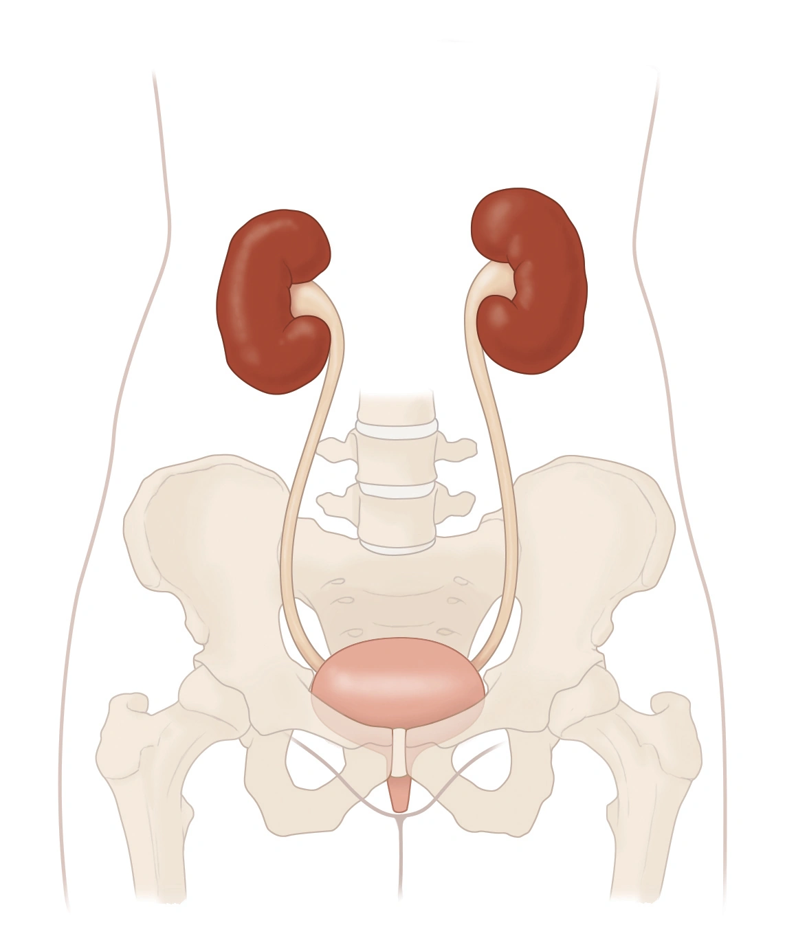
Reading this diagram
The two kidneys are at the top. The ureters lead down to the bladder. Below it, the urethra carries urine out of the body.
For women and men, intradetrusor injections may be offered for selected cases of overactive bladder inadequately controlled by initial treatments. Neurological indications require an appropriate assessment.
The medicine is injected into the bladder muscle under endoscopic guidance. Effects are temporary; infection and retention risks, possible need for self-catheterisation, and bladder-emptying follow-up are explained.
In a surgical clinic · when indicatedCystocele & pelvic organ prolapseAssessing symptoms and pelvic organ support.
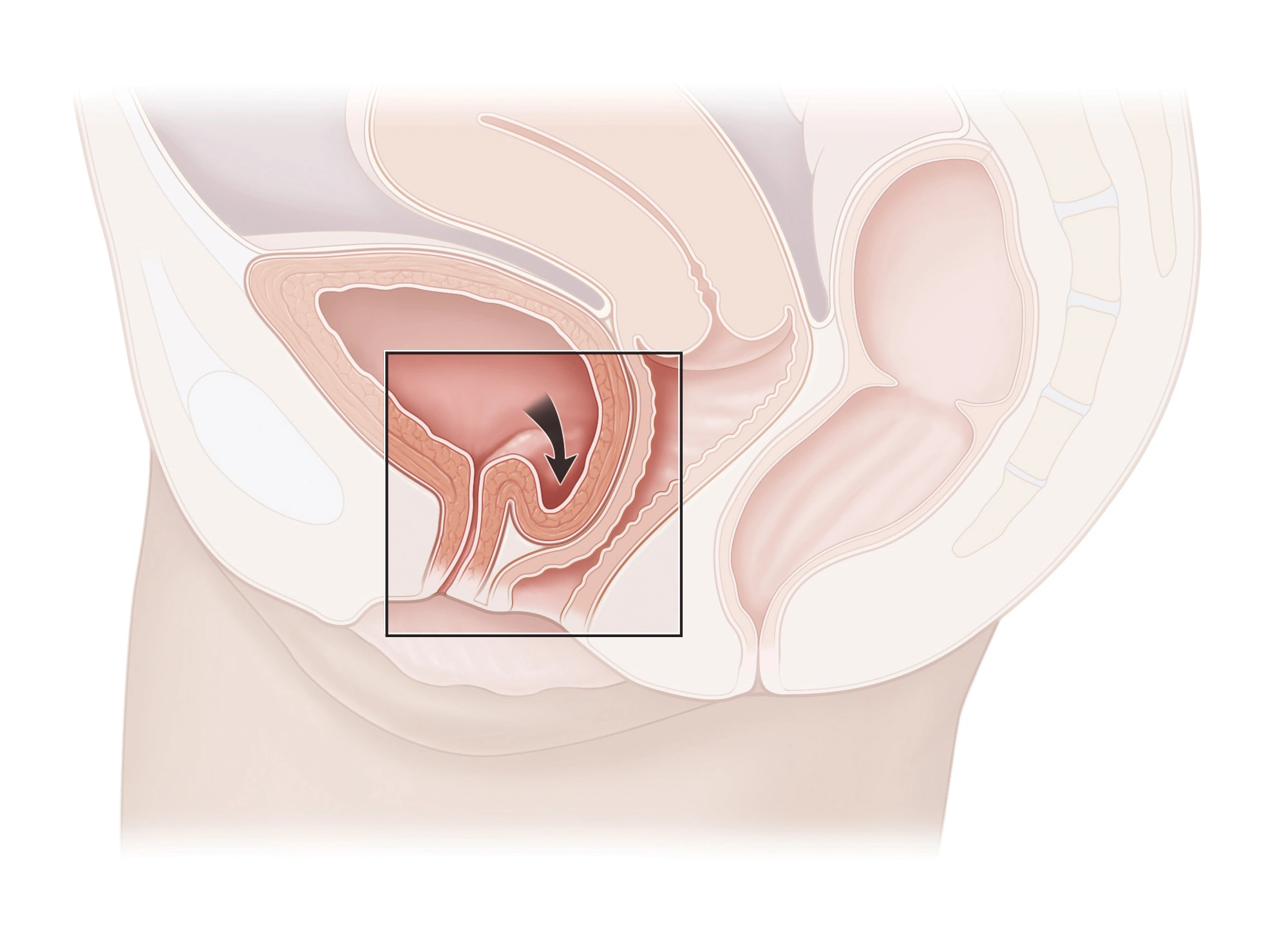
Reading this diagram
The boxed area and arrow show the bladder bulging into the vaginal wall. The drawing depicts a cystocele.
Assessment of prolapse, urinary symptoms and personal goals. Options include pelvic floor therapy, a pessary or surgery according to the findings. Laparoscopic sacrocolpopexy is an option for selected prolapse, performed in a surgical clinic.
Selection depends on the compartments involved and the patient’s plans. Benefits, risks, recurrence and implant-specific complications where relevant are discussed.
In a surgical clinic · when indicatedStress incontinence: TOT / TVT slingsA different indication from prolapse repair.
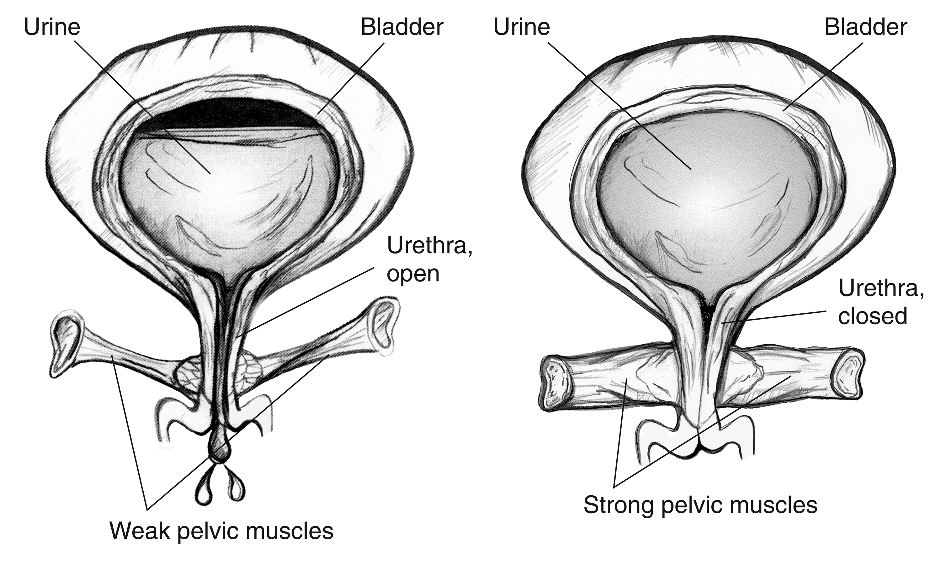
Mid-urethral slings may treat selected stress urinary leakage after assessment and discussion of options, including pelvic floor therapy. TVT uses a retropubic route; TOT a transobturator route.
Selection is individualised for surgery in a clinic. Pain, voiding difficulties and sling-related complications are explained. These procedures alone do not repair a cystocele or prolapse.
Urological oncology
Prostate, bladder and kidney
Consultation & assessmentEarly detection of prostate cancerA discussion based on age and individual risk.
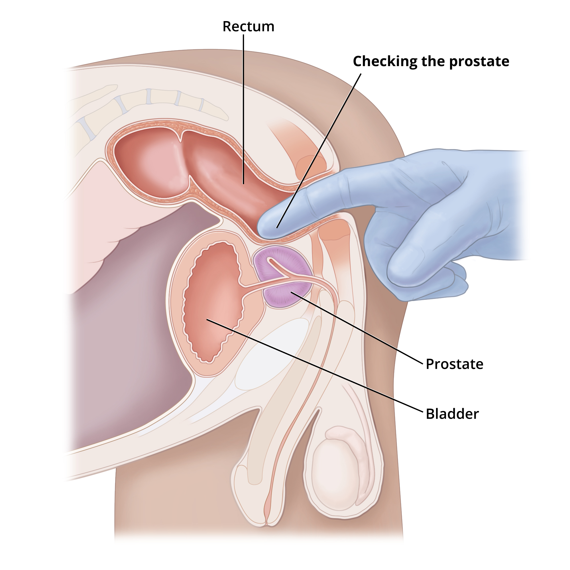
For men without symptoms, discussion may begin around age 50 without particular risk factors, or around 45 with a family history, especially a relative diagnosed before age 60. Certain genetic risks warrant earlier assessment.
Assessment centres on PSA (prostate-specific antigen), with digital rectal examination according to the situation. Benefits, limitations and overdiagnosis are discussed. Testing intervals are individualised; annual PSA and rectal examination are not a universal rule.
Coordinated care · specialist institutionLocalised prostate cancerChoosing a strategy according to the risk level.
Reading this diagram
The prostate sits below the bladder. The urethra passes through the prostate and then the penis. The rectum lies behind them.
Discussion of assessment, active surveillance when appropriate, or local treatment. Open or robot-assisted surgery is arranged in a suitably equipped institution, according to the indication and operating team.
Radiotherapy, sometimes combined with hormone therapy, is coordinated with specialists. Chemotherapy is not a routine treatment for localised prostate cancer.
Practice follow-up & oncology coordinationAdvanced cancer, hormone therapy & resistanceRegular clinical and laboratory follow-up.
Reading this diagram
The prostate sits below the bladder. The urethra passes through the prostate and then the penis. The rectum lies behind them.
Metastatic prostate cancer follow-up includes consultations, laboratory tests and imaging according to disease progression. Hormone injections may be given every three months, depending on the medicine and prescription.
Care includes monitoring bone, metabolic, cardiovascular and sexual effects. Treatment is not always limited to hormone therapy alone: combination treatments and castration-resistant disease are discussed with the oncology team.
At the practice · when indicatedBladder tumours & intravesical BCGRisk-based care after tumour resection.
Reading this diagram
The two kidneys are at the top. The ureters lead down to the bladder. Below it, the urethra carries urine out of the body.
After endoscopic resection and pathology review, BCG treatment at the practice may be indicated for selected non-muscle-invasive bladder tumours, particularly high-risk disease. It is not suitable for every bladder tumour.
Induction, maintenance and surveillance are adapted to EAU and AUA/SUO guidance, tolerance and tumour risk. Safety is checked before each instillation, particularly if there is symptomatic urinary infection or visible blood in the urine. Very high-risk or resistant disease needs specialist reassessment.
Coordinated care · specialist institutionDetection and assessment of kidney tumoursFrom a renal mass to a treatment strategy.
Assessment of an imaging abnormality or suggestive symptoms, characterisation of the mass and staging as appropriate.
Surgical strategy and other options are discussed according to the tumour and kidney function, with care arranged in a suitable institution. This does not imply routine population-wide kidney cancer screening.
In a surgical clinic · when indicatedEndoscopic resection of bladder tumoursRemoving the lesion and obtaining tissue for analysis.
Reading this diagram
The two kidneys are at the top. The ureters lead down to the bladder. Below it, the urethra carries urine out of the body.
Transurethral resection of bladder tumour (TURBT) in a surgical clinic. Pathology establishes tumour grade and depth of involvement, guiding further treatment.
Conventional resection or en-bloc resection in selected cases depends on the lesion and available equipment. Resection and specimen quality are central; repeat resection or additional treatment may be needed following the results.
Consultation & assessmentAssessment, prevention & medical treatmentIdentifying the stone and contributing factors.
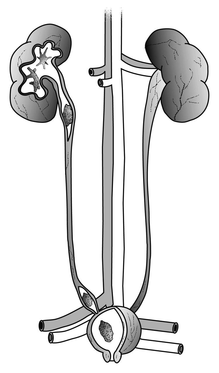
Reading this diagram
The cutaway areas show possible stone locations: in the kidney, the ureter and the bladder.
Localisation and assessment of stones, analysis of retrieved stone composition and metabolic evaluation when appropriate. Prevention advice is individualised.
Some stones can be monitored or managed medically to assist passage when appropriate. Dissolution mainly applies to selected uric acid stones under supervision; not all stones can be dissolved.
Coordinated care · specialist institutionLithotripsy, laser endoscopy & percutaneous surgerySelecting the approach for the particular stone.
Reading this diagram
The cutaway areas show possible stone locations: in the kidney, the ureter and the bladder.
Options include extracorporeal shock wave lithotripsy, ureteroscopy with laser fragmentation and percutaneous nephrolithotomy with an appropriate fragmentation technique, including laser.
Selection depends on stone size, location, composition and the patient’s condition. These procedures are arranged in appropriately equipped facilities. Pain with fever or inability to pass urine requires urgent assessment.
In a surgical clinic · when indicatedSemi-rigid or flexible ureteroscopy & laserTreating ureteric and kidney stones through the urinary tract.
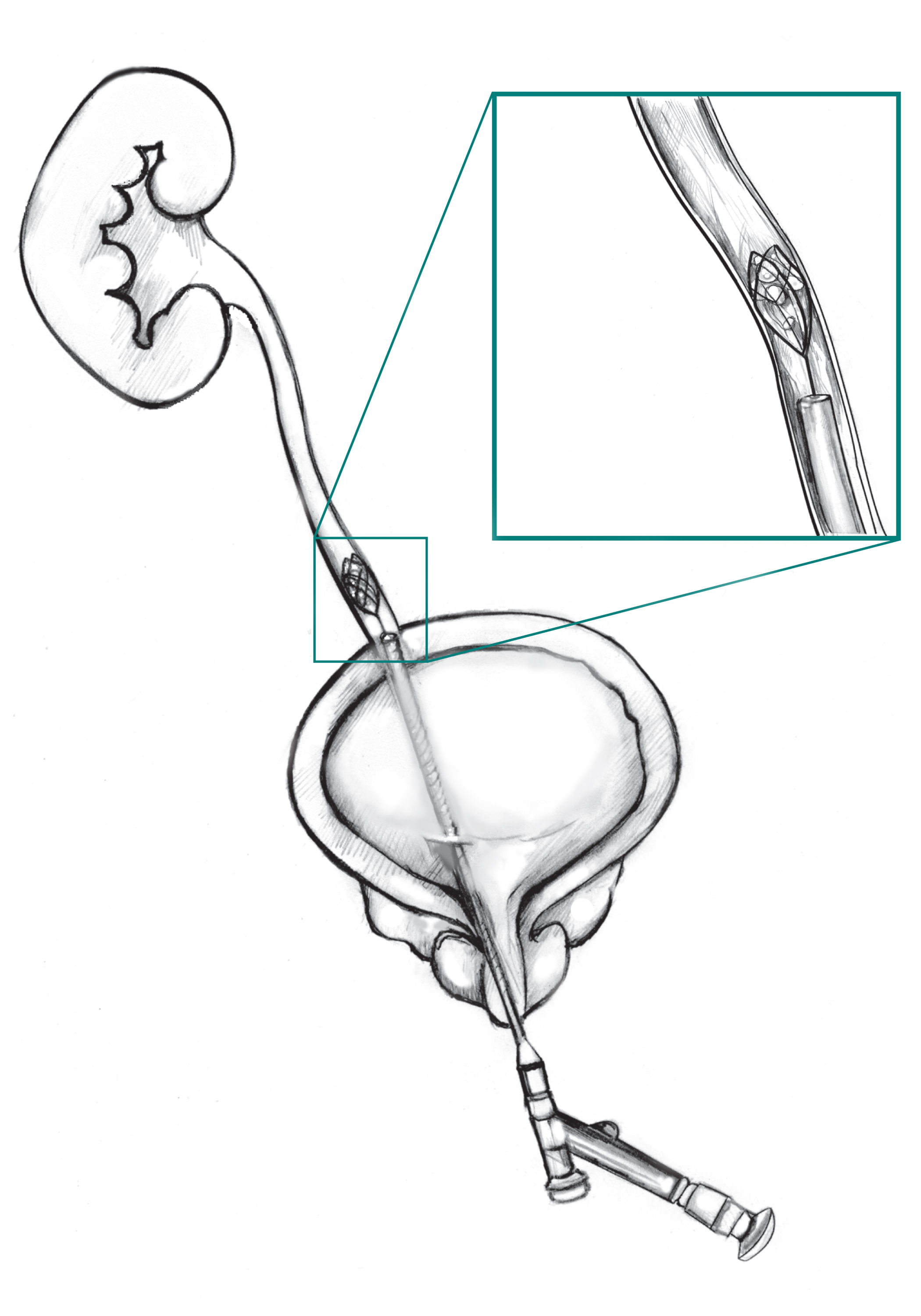
Semi-rigid ureteroscopy is used for selected ureteric stones. Flexible ureteroscopy reaches the kidney collecting system for laser stone fragmentation. Choice depends on location, size and anatomy.
The procedure is performed in a surgical clinic following assessment. A temporary ureteric stent or staged procedures may be needed; their purpose and follow-up are explained.
Consultation & assessmentSexually transmitted infectionsConfidential care for adults of all ages.

Assessment of symptoms and exposures, appropriate sampling, treatment and follow-up. Depending on the situation, testing for other infections, including HIV and syphilis, is offered.
Prevention advice and partner management are discussed according to the infection, to reduce transmission and reinfection.
Andrology & fertility
Male assessment and testicular sperm retrieval

Consultation & assessmentMale infertility assessment & varicoceleLooking for causes within the couple’s overall assessment.
Medical history, genital examination, review of semen analysis and hormonal or other investigations when indicated. A varicocele is assessed and treatment discussed when it may contribute to fertility difficulties.
Male assessment forms part of the couple’s evaluation and, when needed, coordination with an assisted reproduction centre.
Coordinated care · specialist institutionAzoospermia & microsurgical sperm retrievalSpecific indications following a full assessment.
Reading this diagram
The prostate sits below the bladder. The urethra passes through the prostate and then the penis. The rectum lies behind them.
Absence of sperm in the ejaculate must be confirmed and its cause investigated. Hormonal and sometimes genetic assessment precedes a retrieval decision.
Microdissection testicular sperm extraction (micro-TESE) may be offered for selected non-obstructive azoospermia, in an operating facility and with the reproduction team. Sperm retrieval cannot be guaranteed.
Consultation & assessmentHydrocele, varicocele & scrotal conditionsAssessing pain, discomfort or swelling.
Examination of the testes and scrotum, with ultrasound when needed. Monitoring or surgery depends on the cause, symptoms and impact, including on fertility.
Sudden severe testicular pain may indicate torsion, a surgical emergency. Go to an emergency department immediately rather than waiting for a scheduled consultation or WhatsApp reply.
Coordinated care · specialist institutionPenile & external genital surgeryIndividual functional and aesthetic assessment.
Reading this diagram
The prostate sits below the bladder. The urethra passes through the prostate and then the penis. The rectum lies behind them.
Consultation to clarify the concern, examine the anatomy and discuss reconstructive or aesthetic surgical options.
Indications, alternatives, limitations and risks are explained. If surgery is selected, it is arranged in a suitable institution; aesthetic or functional outcomes cannot be guaranteed.
At the practice · selected patientsCircumcision for childrenPlanned following a preliminary consultation.
Reading this diagram
The prostate sits below the bladder. The urethra passes through the prostate and then the penis. The rectum lies behind them.
Circumcision at the practice for children whose age, health and care requirements allow it. Prior examination, parental information and consent, and appropriate anaesthesia are essential. A surgical clinic is recommended when needed.
Conditions such as hypospadias, local infection or bleeding disorders require specific assessment before proceeding. Aftercare and postoperative review are explained.
At the practice · selected patientsVasectomy under local anaesthesiaPermanent male contraception through a small opening.
Reading this diagram
The prostate sits below the bladder. The urethra passes through the prostate and then the penis. The rectum lies behind them.
Vasectomy through a micro-incision at the practice for selected adults following counselling and informed consent. Permanence, alternatives and possible complications are discussed before deciding.
It is not immediately effective: another contraceptive method is needed until the doctor confirms clearance following postoperative semen testing. Vasectomy does not protect against STIs.
In a surgical clinic · when indicatedInguinal & inguinoscrotal herniasAssessment and surgical repair in a clinic.
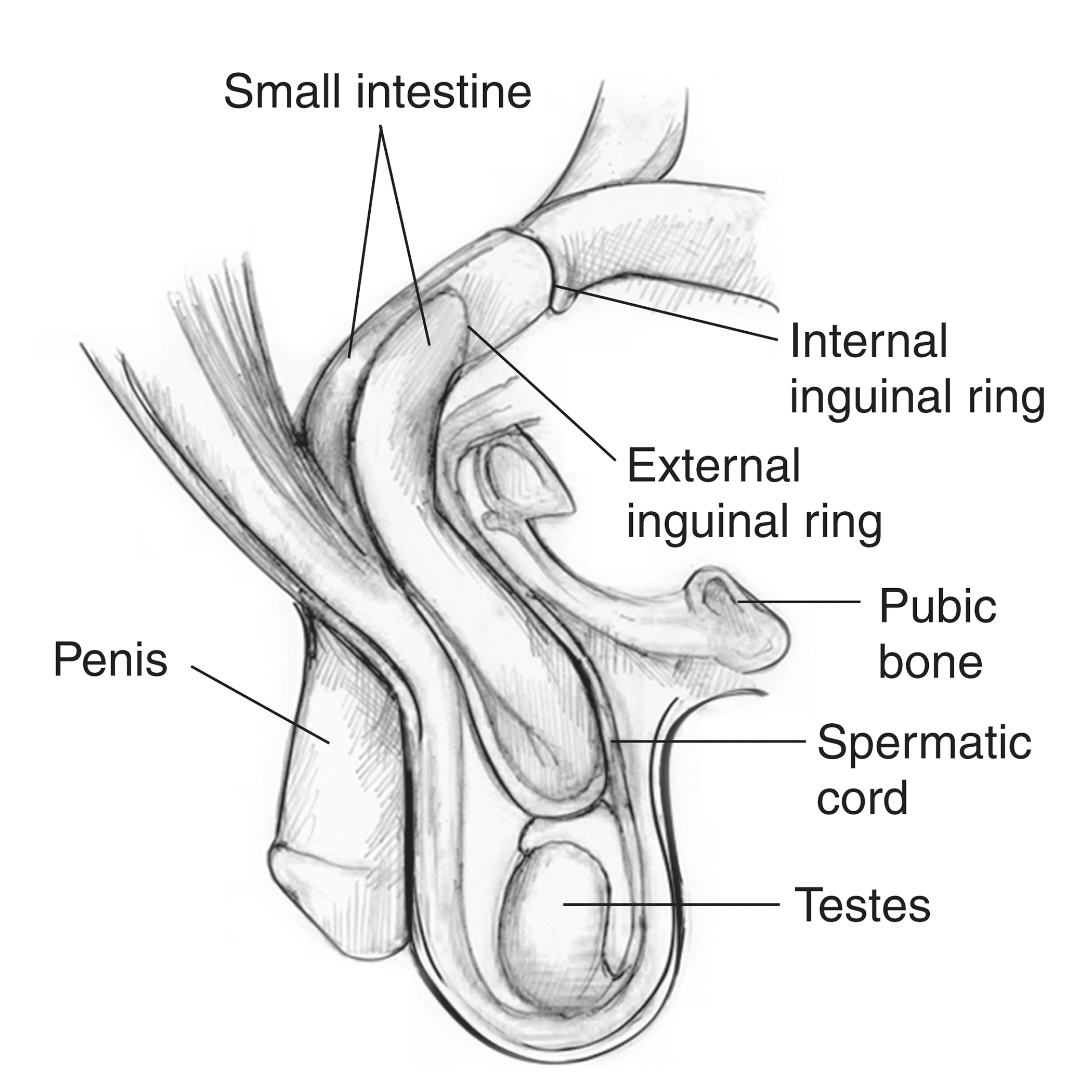
Assessment of a groin swelling or a swelling extending into the scrotum, pain and its impact. Repair and technique are discussed according to the hernia, the patient’s health and the surgical team.
A painful lump that no longer reduces, especially with vomiting, requires immediate emergency assessment.
Emergency · surgical clinic or hospitalPenile fractureAn emergency requiring a surgical facility.
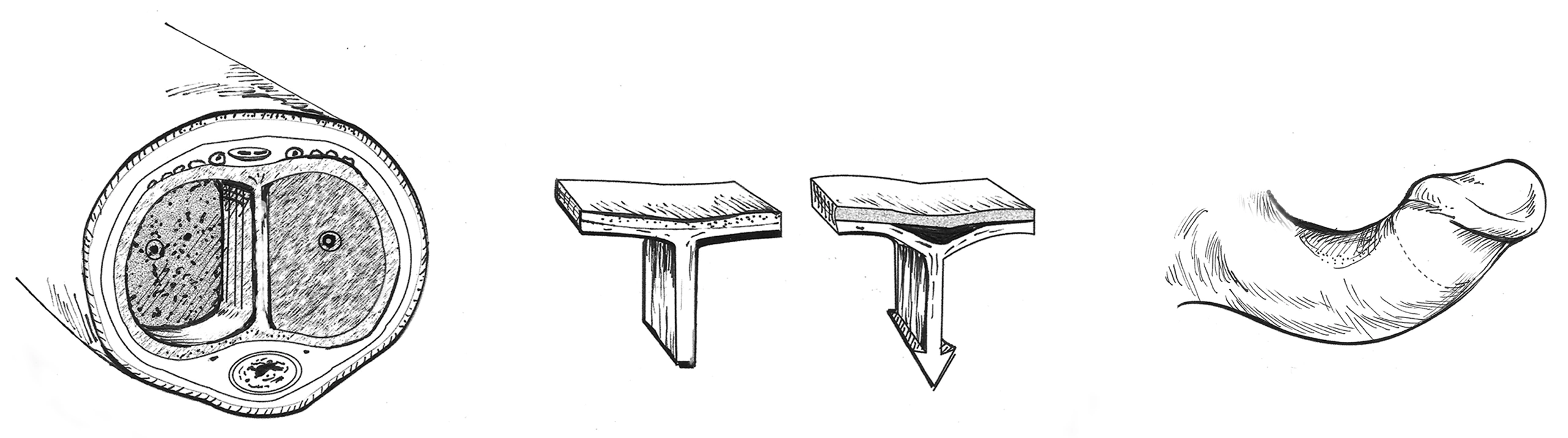
After injury to an erect penis, a cracking sound, pain, rapid loss of erection or marked swelling may suggest a tear in the covering of the erectile bodies.
Urgent assessment in a surgical clinic or hospital also checks for associated urethral injury. Prompt surgical repair is recommended when a fracture is confirmed. Do not delay seeking care while waiting for the practice to reply.
Sexual health
Erectile dysfunction and sexological care
Consultation & assessmentOrganic erectile dysfunctionAssessment before treatment selection.
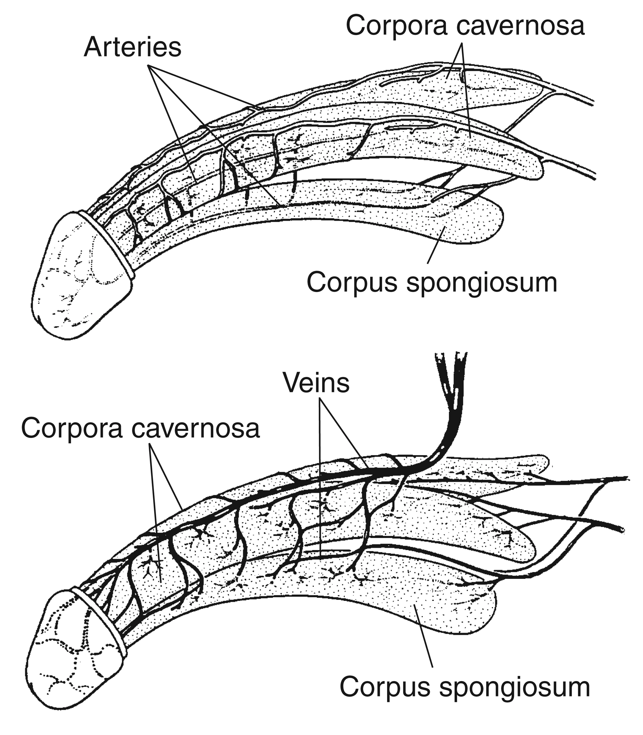
Evaluation of vascular, metabolic, hormonal, neurological, medication-related and psychological factors. Several causes may coexist.
Care takes account of overall health, expectations and contraindications. Established treatments and sexological support are discussed before considering complementary techniques.
Selected indication · limited evidenceLow-intensity shock wave therapyAn option for selected vascular profiles.
This approach may be discussed for selected vasculogenic erectile dysfunction after assessment and counselling about generally modest, variable benefits. Indications and protocols depend on the patient.
Guidelines differ: the EAU gives a weak recommendation for selected patients, while the AUA considers it investigational. Radial waves are not equivalent to the studied focused shock waves; success is not guaranteed.
Information · clinical researchIntracavernous PRP: an experimental approachUnderstanding the current evidence.
Platelet-rich plasma injections into the corpora cavernosa are being studied for erectile dysfunction. Efficacy, optimal protocols and long-term safety are not sufficiently established.
PRP is not presented as an established standard treatment: the EAU and AUA restrict this approach to clinical research. Consultation allows discussion of this uncertainty and recognised alternatives.
At the practice · when indicatedLearning to use a vacuum erection devicePractical guidance with an appropriate medical device.
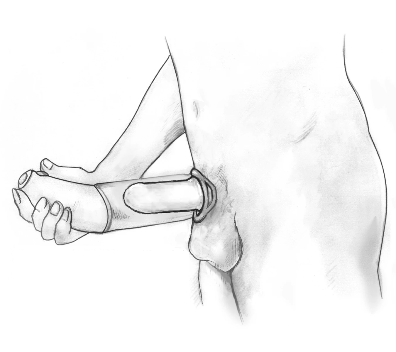
Sessions at the practice to learn how to use a medical vacuum device, reviewing handling, comfort and use instructions with the patient.
If a constriction ring is used, it must not remain in place for more than 30 minutes. Tell the doctor about any bleeding disorder or anticoagulant use before choosing this option.
At the practice · when indicatedLearning intracavernosal self-injectionPrescribed alprostadil / prostaglandin E1.
Following erectile dysfunction assessment, the doctor determines the dose and supervises initial use at the practice. Home use becomes possible once the patient has mastered the technique and instructions.
Prescribed frequency, hygiene and what to do if problems occur are explained. An erection persisting for four hours requires immediate emergency care; do not increase the dose yourself.
Selected indication · limited evidencePeyronie’s disease & shockwave therapyClarifying the aim: pain or deformity.
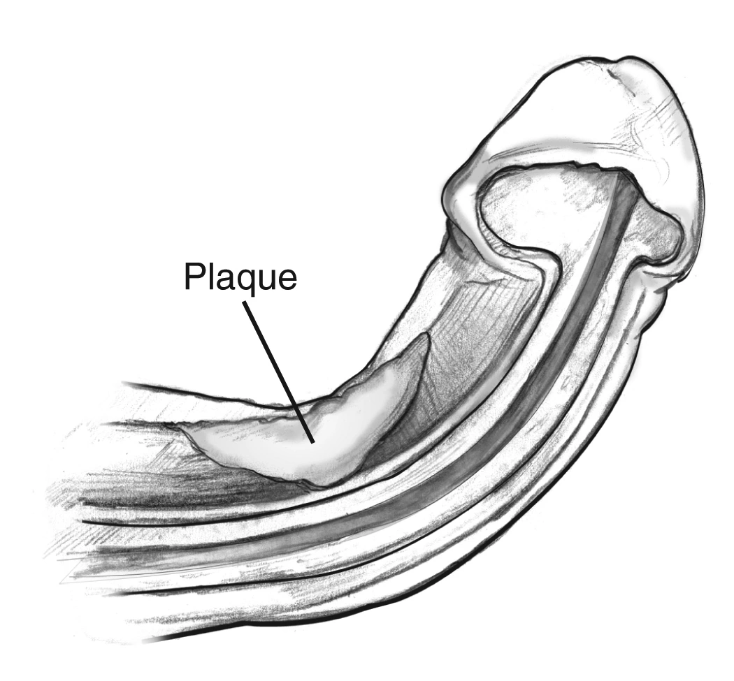
Consultation assesses pain, curvature and its progression, as well as erectile function. Options depend in part on whether the disease is active or stable.
The EAU gives a weak recommendation for appropriate extracorporeal shockwaves to address pain during the active phase. They are not recommended to correct curvature or reduce plaque. No device justifies promising straightening with this technique.
Consultation & assessmentPsychosexual & couples therapySupport for intimate life and relationships.
Individual or couple consultations explore sexual difficulties, their impact and each person’s expectations. Work may focus on communication, performance anxiety and adapting to treatment.
Participation is voluntary and confidentiality is respected. Support can complement medical assessment and, when appropriate, specialist psychological care.
Care & independence
Catheterisation, neurogenic bladder and urinary diversions
Understand the conditionsUrgent · practice or emergency departmentAcute urinary retention & catheterisationSudden inability to pass urine needs prompt assessment.

Assessment and bladder drainage with a catheter at the practice when the clinical situation and facilities allow, together with investigation of the cause. Difficult catheterisation or complications require care in a surgical clinic or emergency department.
Do not wait for a WhatsApp reply or a later appointment if you cannot urinate and are in pain. If the practice cannot assess you immediately, go to an emergency department.
At the practice · when indicatedLearning intermittent self-catheterisationBuilding confidence in safe bladder emptying.
Reading this diagram
The two kidneys are at the top. The ureters lead down to the bladder. Below it, the urethra carries urine out of the body.
Teaching sessions at the practice for patients with neurogenic bladder or another indication for self-catheterisation. Equipment and frequency are individualised, with demonstration and assessment of the patient’s technique.
Training covers hygiene, difficulties and warning signs, alongside bladder and kidney follow-up appropriate to the neurological condition.
At the practice · when indicatedNephrostomy & urinary stoma careChecking the opening, surrounding skin and drainage.
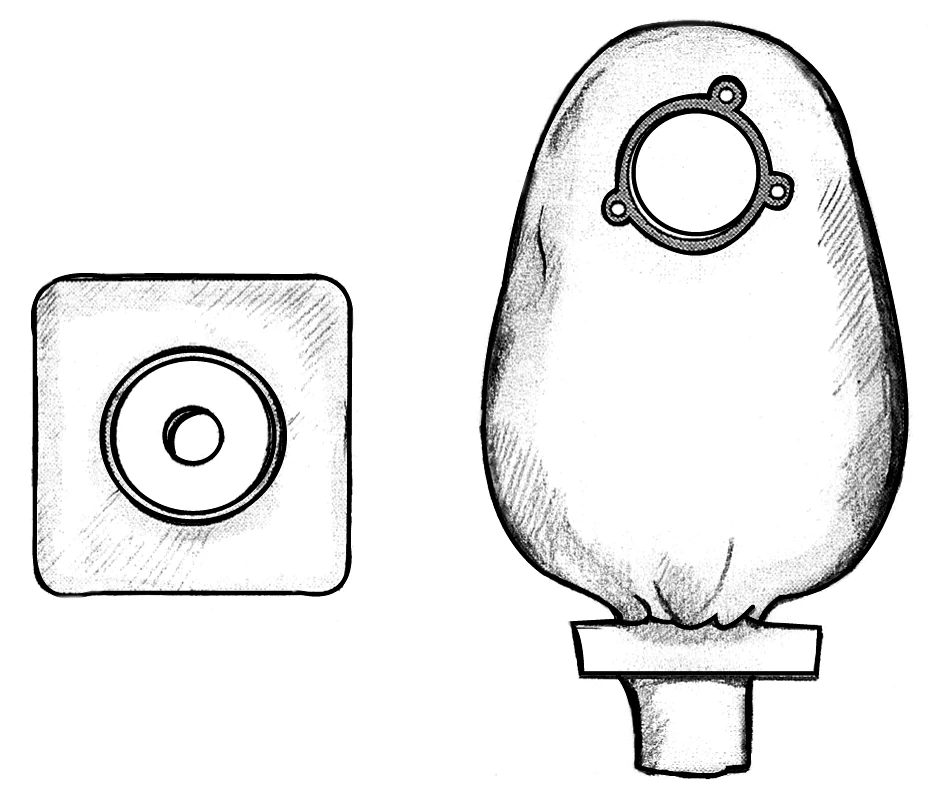
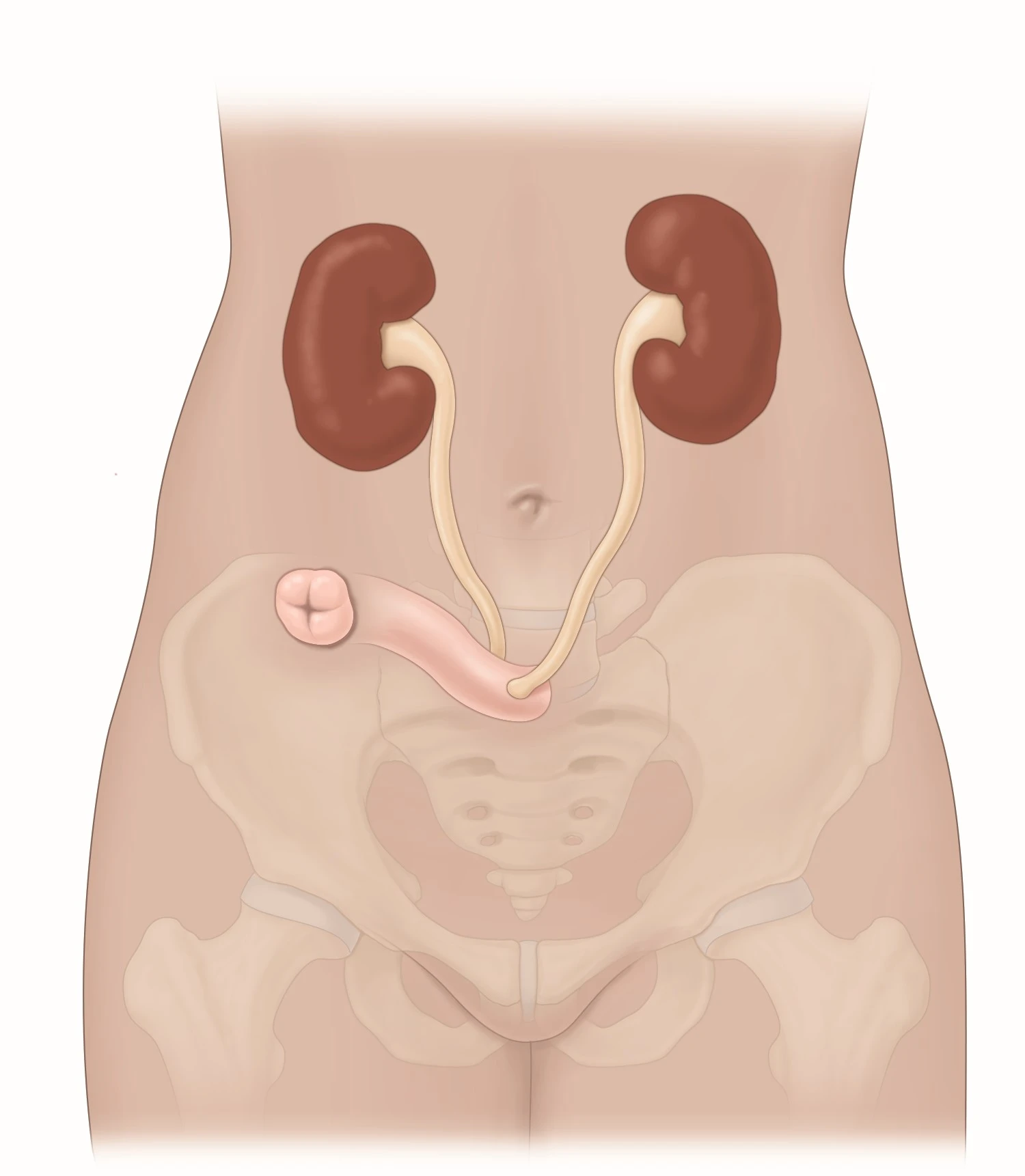
Care, dressings and support for nephrostomies and cutaneous ureterostomies, including after bladder cancer surgery. A nephrostomy drains the kidney directly; a ureterostomy brings a ureter to the skin. Care is tailored to the actual urinary diversion.
Care is coordinated with the surgical and nursing teams. Drain changes requiring specialist facilities are arranged appropriately. Fever, pain or loss of drainage needs prompt advice.
Medical references
Content checked on 3 October 2026. Indications depend on your assessment and evolving clinical guidance.
- EAU · Prostate cancer — early detection
- EAU · Prostate cancer — treatment
- EAU · Non-muscle-invasive bladder cancer
- AUA/SUO · Non-muscle-invasive bladder cancer (2024)
- EAU · Renal cell carcinoma
- EAU · Male lower urinary tract symptoms
- NIDDK · Interstitial cystitis — diagnosis
- EAU · Urolithiasis
- EAU · Male infertility
- EAU · Erectile dysfunction
- AUA · Erectile dysfunction (2018)
- CDC · STI and HIV infection risk assessment
- NHS · Testicular pain and emergencies
- EAU · Penile curvature / Peyronie’s disease
- EAU · Neuro-urology
- EAU · Non-neurogenic female lower urinary tract symptoms
- NICE · Urinary incontinence and pelvic organ prolapse
- EAU · Male lower urinary tract symptoms — treatment
- EAU · Bladder tumour diagnosis and resection
- EAU · Paediatric urology — foreskin conditions
- BAUS · Vasectomy
- EAU · Vacuum erection devices — patient information
- BAUS · Penile self-injection therapy
- EAU · Urological trauma
- NHS · Urinary catheters
- Guy’s and St Thomas’ NHS · Nephrostomy follow-up
- NHS England · Inguinal hernia — decision aid
- EAU · Testicular cancer — diagnostic evaluation
APPOINTMENTS & DIRECTIONS
Let’s talk about
your health.
The secretary can help with appointments and examination preparation instructions.
Before your visit
Opening hours & visits
| Days | Hours |
|---|---|
| Monday to Friday | 9 am – 4 pm · without a midday break |
| Saturday and Sunday | Closed |
Some mornings are spent operating at the clinic, on a variable schedule. Please contact the practice to confirm your appointment and the doctor’s availability before travelling.
Confirm my appointment05 37 40 80 00The practice in Temara
Avenue Hassan II, Résidence Ilham 3Building A, 2nd floor, Office 11
Temara Mall · TemaraFind the practice on Google