
Dr Ali Beddouche
My procedure pathway
Guidance before the procedure, during it and after returning home.
Your team specifies timing, anaesthesia and return to activities.
Cystoscopy
Examine the bladder for bleeding, an abnormality or tumour surveillance.
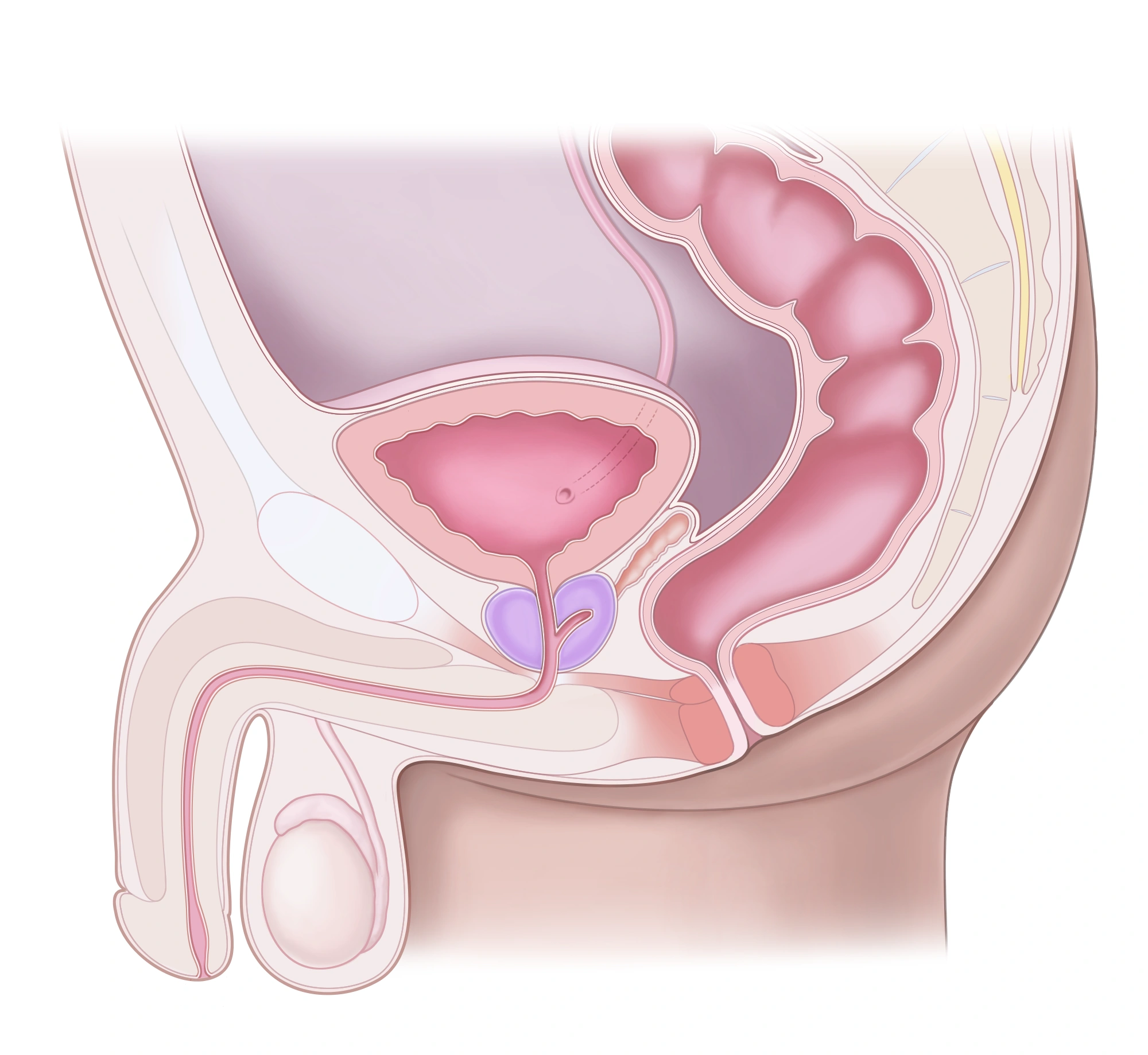
Reading this diagram
The prostate sits below the bladder. The urethra passes through the prostate and then the penis. The rectum lies behind them.
Before
Report infection, medicines and allergies. The team determines urine testing and anaesthesia requirements.
During
A fine camera passes through the urethra. Flexible examination often uses local anaesthetic gel; additional treatment may require another anaesthetic.
After
Mild burning and pink urine may be temporary. Follow discharge advice; fever or inability to urinate requires prompt care.
What to expect
Shows the lining; a biopsy may be needed to identify a lesion.
Risks and limitations
Infection or bleeding can occur. A normal examination does not explain every urinary symptom.
Prostate biopsy
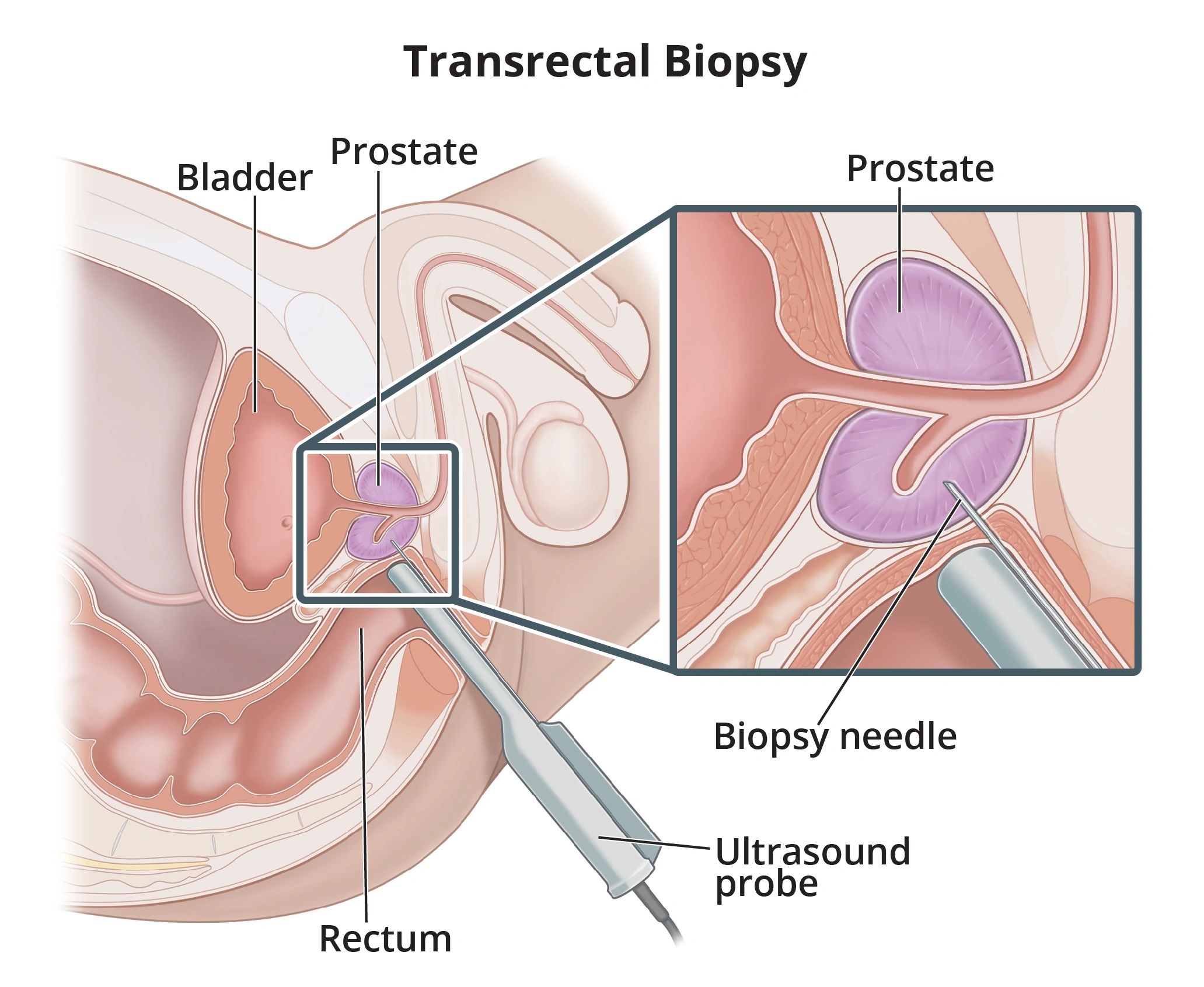
Sample tissue when assessment suggests cancer; PSA alone does not determine the decision.
Before
MRI and individual risk guide sampling. Arrange blood-thinner management and infection prevention with the clinician.
During
Ultrasound guides targeted or systematic samples. A transperineal approach is generally favoured to reduce infection risk.
After
Discuss the pathology report at follow-up. Blood in urine or semen can occur; fever or retention needs urgent assessment.
What to expect
Identifies cancer in sampled tissue and determines its grade.
Risks and limitations
A negative biopsy does not exclude every cancer. Bleeding, infection or retention can occur.
Ureteroscopy and laser
Treat selected ureter or kidney stones according to size, location and effects.
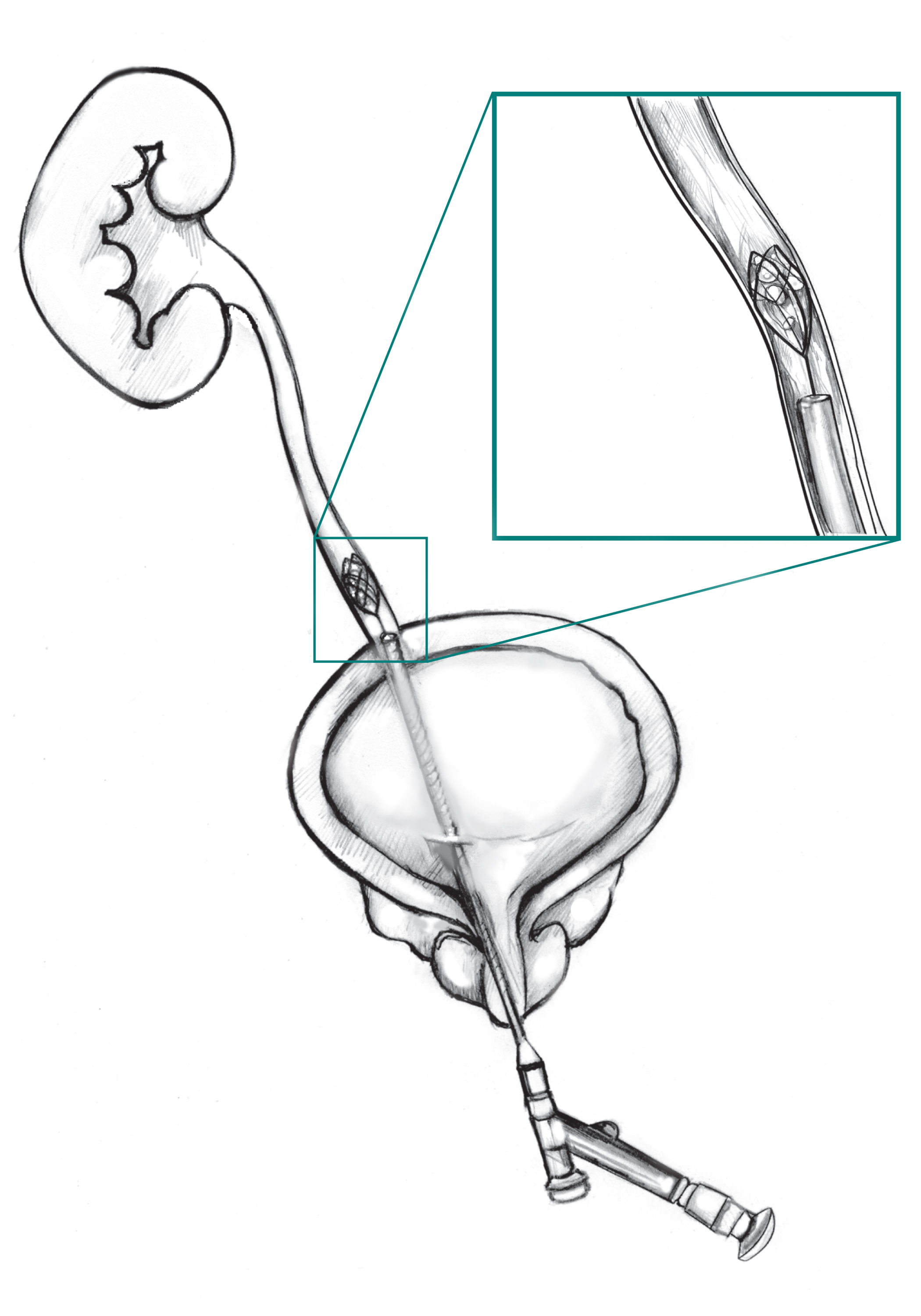
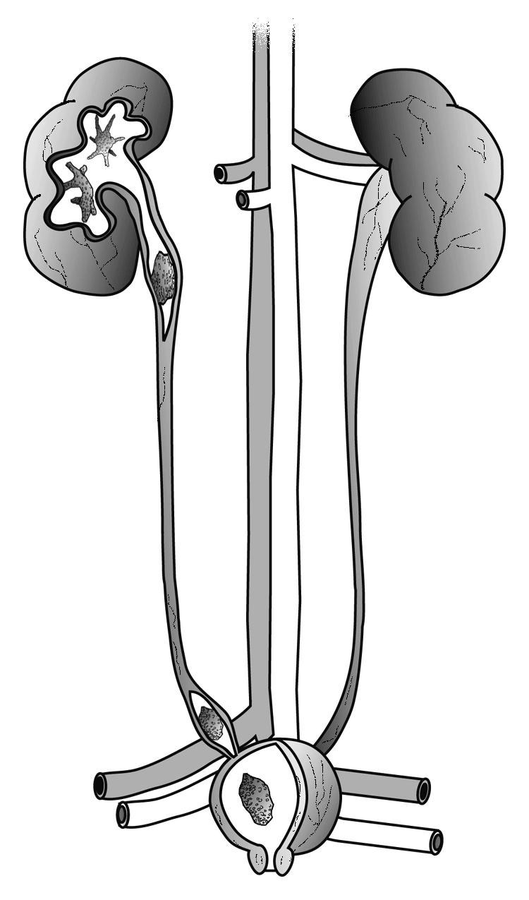
Reading this diagram
The cutaway areas show possible stone locations: in the kidney, the ureter and the bladder.
Before
Imaging and infection assessment; an infected obstruction needs drainage before definitive stone treatment. Follow individual anaesthetic advice.
During
An endoscope enters through the natural urinary passage. The stone is extracted or fragmented, often with a laser; a JJ stent may be placed.
After
Check clearance and arrange stent removal. Stone analysis helps plan recurrence prevention.
What to expect
Can clear the urinary passage; more than one session may be needed.
Risks and limitations
Infection, ureter injury, remaining fragments and stent discomfort are possible.
Shock-wave lithotripsy — SWL
Break suitable stones with shock waves; density, size and location affect selection.
Reading this diagram
The cutaway areas show possible stone locations: in the kidney, the ureter and the bladder.
Before
Locate the stone, assess infection and review medicines that increase bleeding. Pregnancy rules out this technique.
During
Targeted waves from outside the body fragment the stone, with pain relief.
After
Fragments pass in urine. Imaging checks clearance; repeat sessions or endoscopy may be needed.
What to expect
Avoids an incision and may allow stone passage.
Risks and limitations
Colic, blood in urine, infection or blockage by fragments may occur; effectiveness varies.
Percutaneous nephrolithotomy — PCNL
Treat mainly large or complex kidney stones.
Reading this diagram
The cutaway areas show possible stone locations: in the kidney, the ureter and the bladder.

Before
CT imaging, infection and anaesthetic assessment; discuss bleeding risk and current medicines.
During
A small tract through the skin gives access to the kidney to break and remove stones under anaesthesia.
After
Hospital monitoring checks bleeding, infection and remaining stones. Temporary drainage may be needed.
What to expect
Removes a substantial stone burden, sometimes in stages.
Risks and limitations
Potential risks include significant bleeding, infection and injury to nearby organs.
JJ ureteric stent
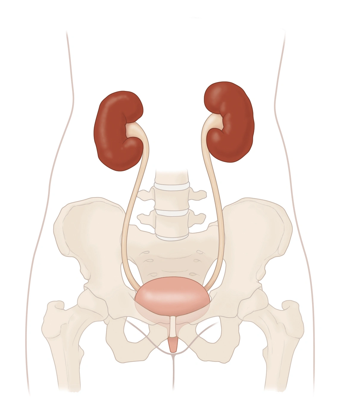
Keep urine flowing from kidney to bladder around an obstruction or after selected procedures.
Reading this diagram
The two kidneys are at the top. The ureters lead down to the bladder. Below it, the urethra carries urine out of the body.
Before
Clarify the reason, possible infection and planned removal or exchange date.
During
An internal tube with curled ends is placed endoscopically, sometimes through kidney access.
After
A removal or exchange schedule is essential. Urgency and flank discomfort may occur.
What to expect
Provides drainage; it does not always treat the cause of obstruction.
Risks and limitations
Infection, migration and encrustation, especially if forgotten. Fever or severe pain requires prompt assessment.
Nephrostomy
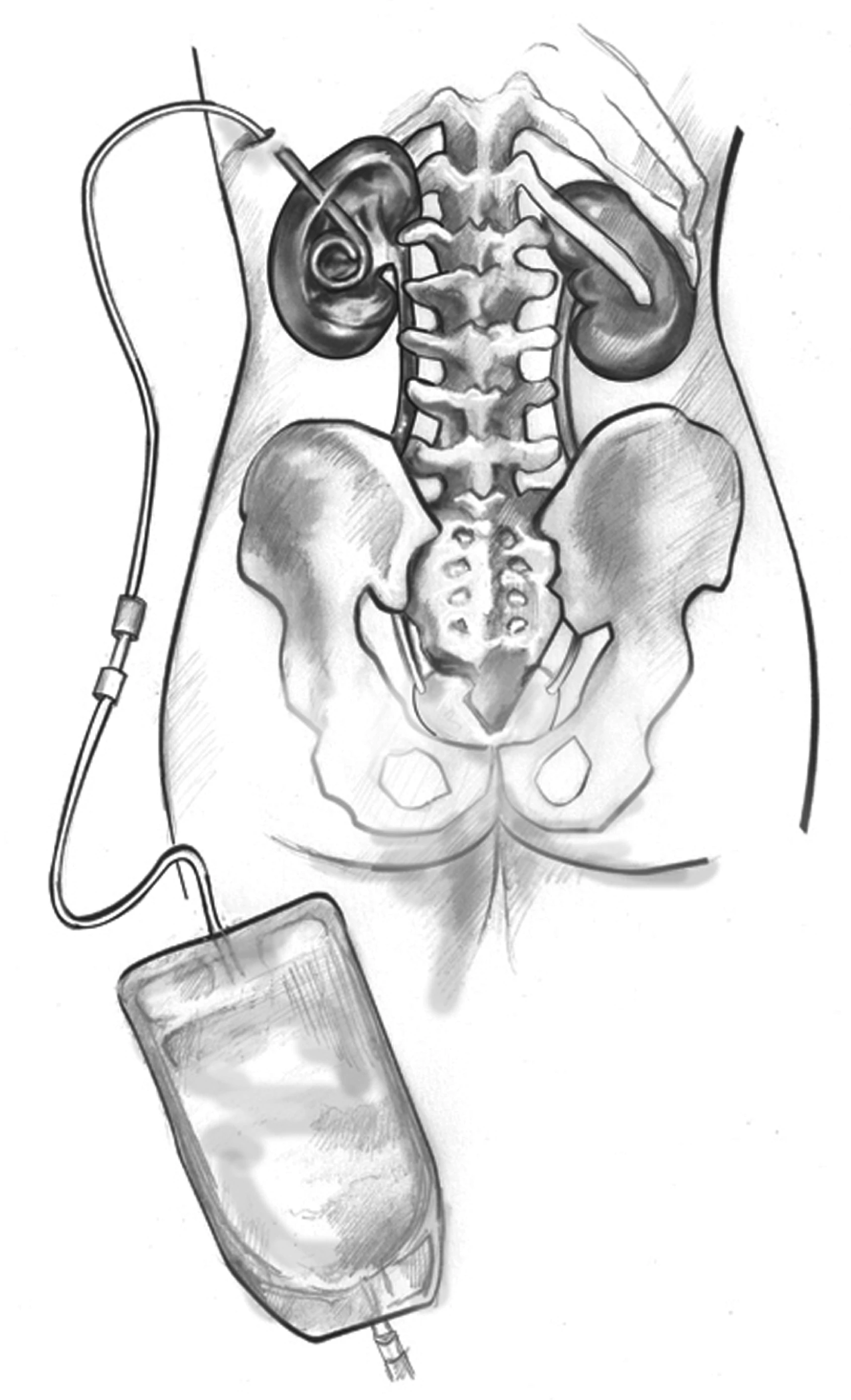
Drain an obstructed kidney, especially urgently when infection is present.
Before
Imaging, clotting assessment and infection treatment are arranged according to urgency.
During
A catheter passes through the skin into the kidney collecting system under imaging guidance and appropriate anaesthesia.
After
Follow advice about the bag, fixation and output. Report fever, displacement or stopped drainage.
What to expect
Relieves pressure while the underlying cause is addressed.
Risks and limitations
Bleeding, infection, displacement or blockage can occur. Do not manipulate or flush without team instructions.
Transurethral resection of the prostate — TURP
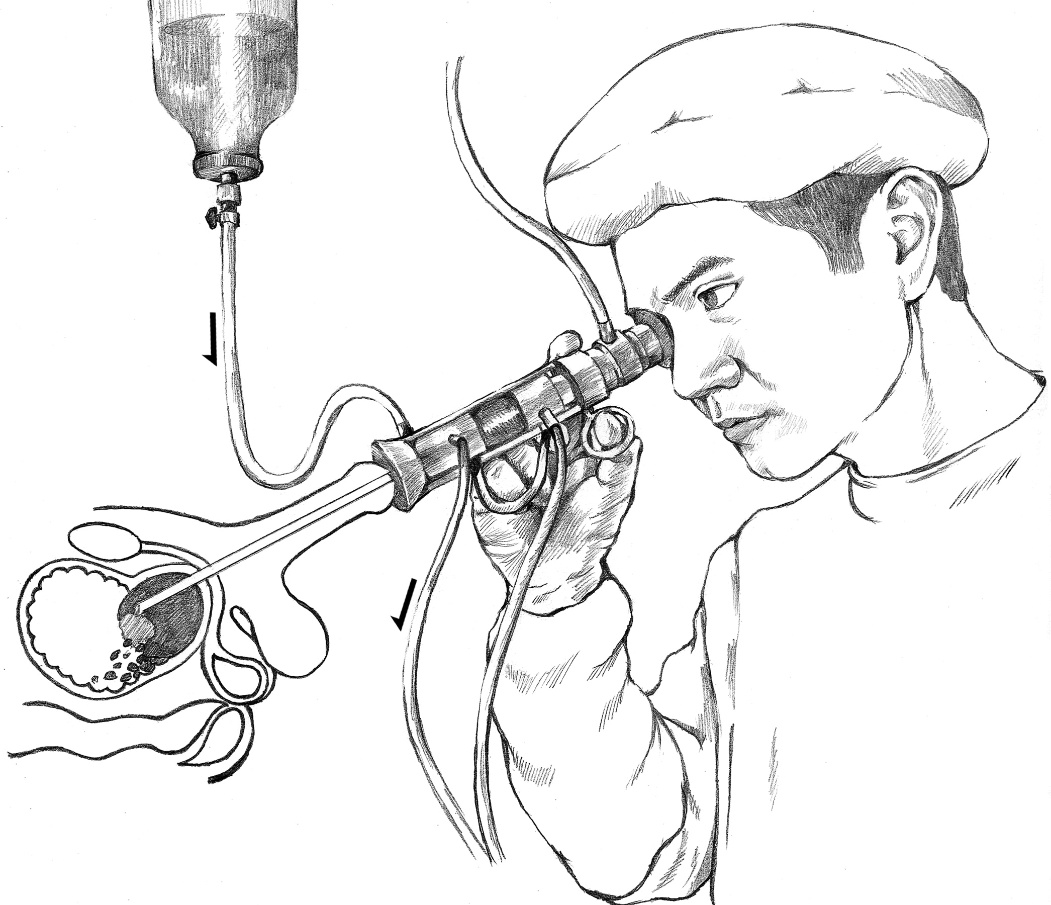
Relieve troublesome or complicated benign obstruction when appropriate.
Before
Urine and anaesthetic assessment, prostate volume and medicine review. Discuss fertility and ejaculation.
During
An endoscope removes obstructing prostate tissue; the whole prostate is not removed.
After
Temporary catheter, gradual return to activity and urination review.
What to expect
Often improves flow; frequency may persist if the bladder also contributes.
Risks and limitations
Retrograde ejaculation is common; bleeding, infection, narrowing or incontinence can occur.
Holmium laser enucleation — HoLEP
Treat benign obstruction, including selected large prostates, depending on expertise and individual needs.
Reading this diagram
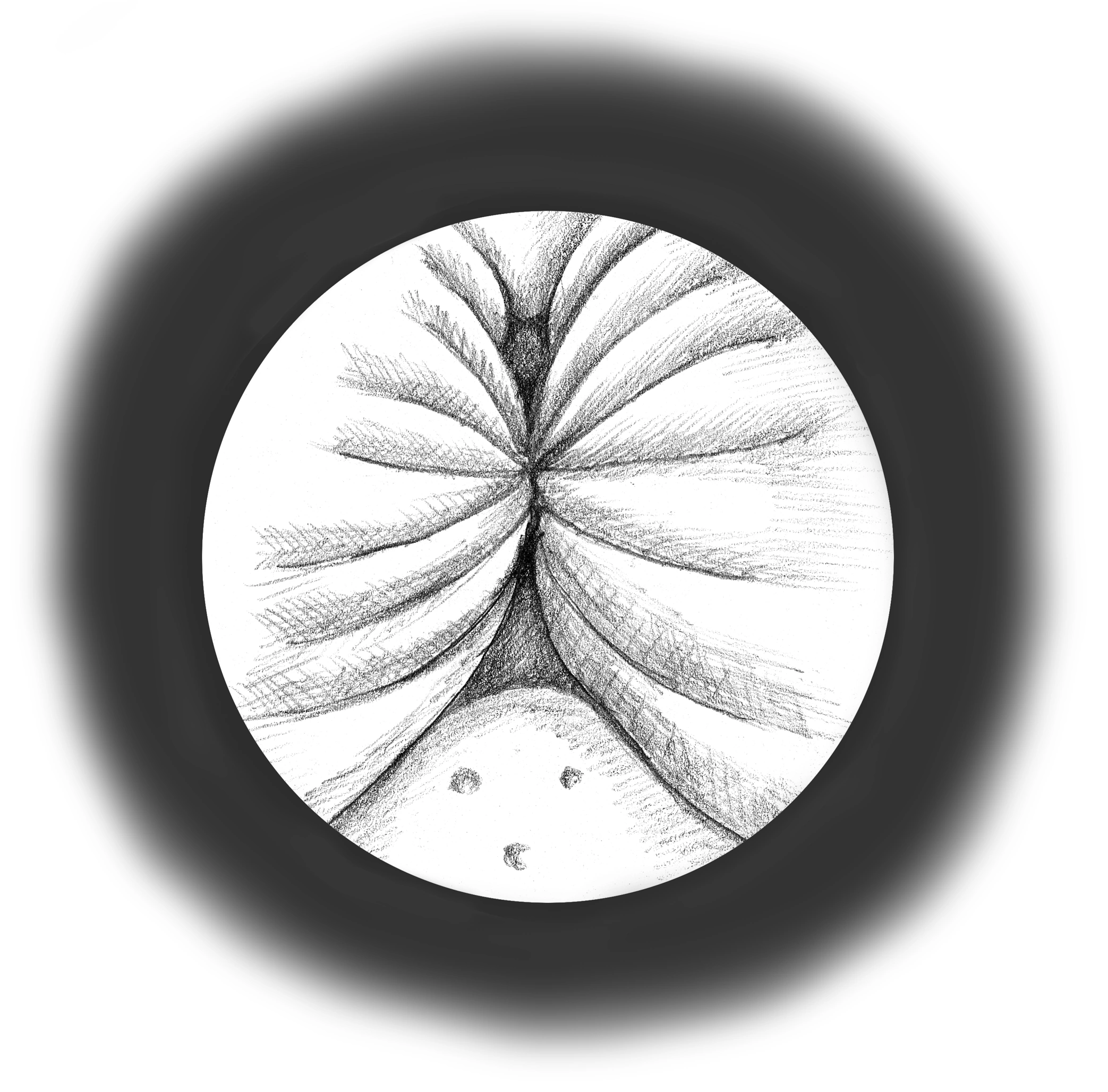
This view looks from inside the urethra. The walls are pushed together, leaving a narrower passage for urine.
Reading this diagram
The prostate sits below the bladder. The urethra passes through the prostate and then the penis. The rectum lies behind them.
Before
Obstruction assessment, urine testing and anaesthetic review. Discuss ejaculation changes.
During
A laser separates the adenoma; tissue is removed through the urethra and examined.
After
Temporary catheter and flow review. Leakage or urgency can accompany recovery.
What to expect
Often provides lasting relief; bladder function also influences the result.
Risks and limitations
Retrograde ejaculation is common; temporary leakage, infection, bleeding or narrowing can occur.
Transurethral bladder tumour resection — TURBT
Remove a bladder lesion and determine its type and depth.
Reading this diagram
The two kidneys are at the top. The ureters lead down to the bladder. Below it, the urethra carries urine out of the body.
Before
Urine assessment, medicine review and anaesthesia; imaging depends on context.
During
An endoscope through the urethra removes tumour tissue for analysis without an abdominal incision.
After
Discuss pathology, possible repeat resection and intravesical treatment; surveillance cystoscopies are needed.
What to expect
Establishes diagnosis and treats selected superficial tumours.
Risks and limitations
Recurrence remains possible. Bleeding, infection or bladder perforation may occur.
Bladder instillations: BCG or chemotherapy
Reduce recurrence or progression of selected non-muscle-invasive tumours according to risk.
Reading this diagram
The two kidneys are at the top. The ureters lead down to the bladder. Below it, the urethra carries urine out of the body.
Before
Check healing, absence of symptomatic infection and product-specific contraindications.
During
Medicine is placed in the bladder through a catheter on a defined schedule.
After
Follow the centre’s hygiene instructions and continue cystoscopic surveillance.
What to expect
Reduces tumour risk when indicated, without eliminating it.
Risks and limitations
Irritation and fever may occur; persistent fever or illness after BCG needs urgent medical contact.
Radical prostatectomy
Treat selected prostate cancers within a risk-based discussion of alternatives.
Reading this diagram
The prostate sits below the bladder. The urethra passes through the prostate and then the penis. The rectum lies behind them.
Before
Appropriate staging, anaesthetic review and counselling about continence, erections and fertility.
During
The prostate and seminal vesicles are removed through open or minimally invasive surgery, sometimes robot-assisted.
After
Temporary catheter, pathology and PSA follow-up; rehabilitation and sexual support as needed.
What to expect
Aims to control cancer; additional treatment may still be required.
Risks and limitations
Incontinence, erectile dysfunction and loss of ejaculation; robotic assistance does not remove these risks.
Partial or radical nephrectomy
Remove a kidney tumour when surgery is indicated; preserve kidney tissue when feasible and appropriate.
Before
Imaging, kidney function and assessment of the other kidney; discuss surveillance or alternatives depending on the lesion.
During
The tumour alone or the kidney is removed through an approach selected for anatomy and expertise.
After
Review kidney function and pathology, with appropriate cancer surveillance.
What to expect
Aims for tumour control while preserving kidney function where possible.
Risks and limitations
Bleeding, urine leakage after partial surgery and reduced kidney function; cure cannot be guaranteed individually.
Cystectomy and urinary diversion
Treat selected invasive bladder cancers or very high-risk disease.
Before
Full assessment, nutrition and anaesthesia; choose and prepare for diversion care. Discuss sexual and reproductive effects.
During
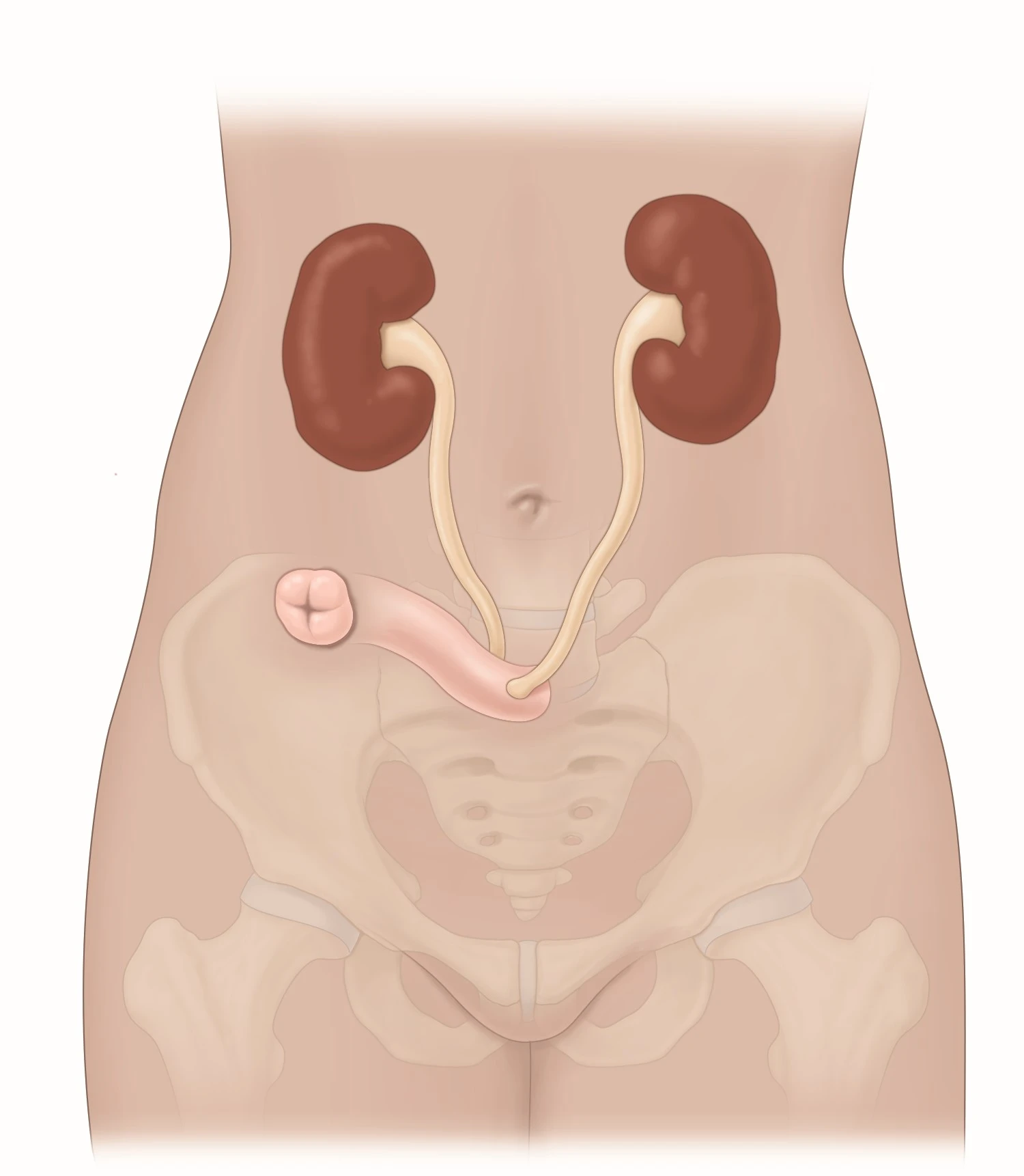
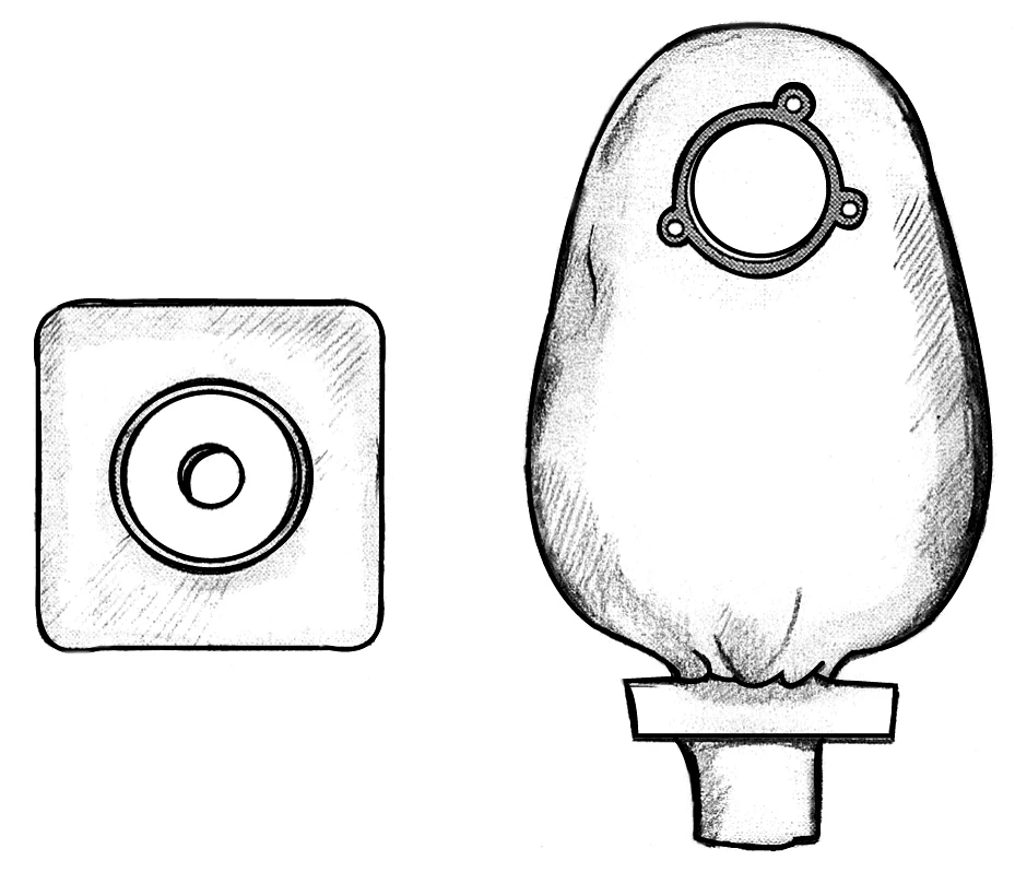
The bladder is removed and a new urine outlet is made, through a stoma or a reservoir in selected cases.
After
Rehabilitation, practical care training and long-term kidney, metabolic and cancer follow-up.
What to expect
Aims to control cancer with a new way to drain urine.
Risks and limitations
Major surgery: infection, thrombosis, bowel complications, sexual changes and diversion-related complications.
Pyeloplasty
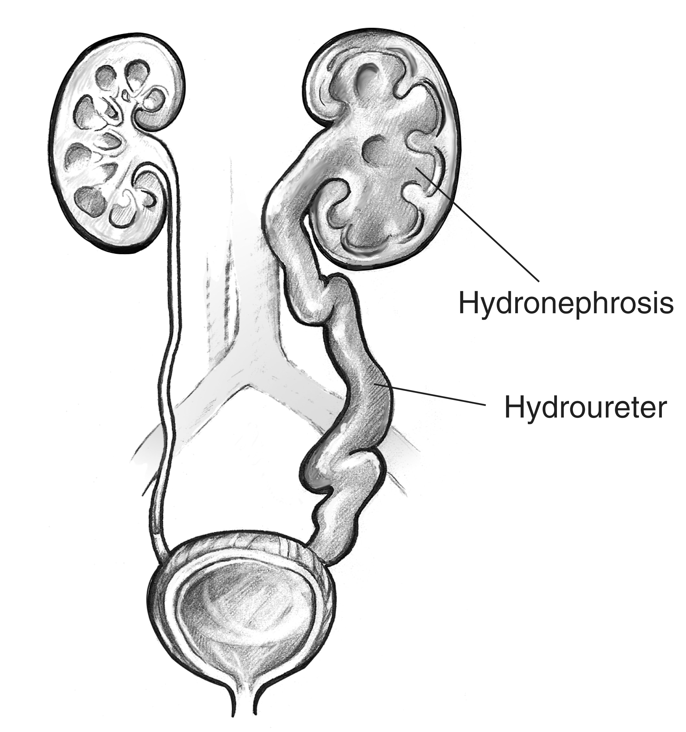
Correct selected narrowings between the renal pelvis and ureter that impair kidney drainage.
Before
Ultrasound, imaging and sometimes a renal scan assess obstruction and function.
During
The junction is reconstructed by open or minimally invasive surgery; a stent may support healing.
After
Planned stent removal and drainage and function checks.
What to expect
Improves drainage; a damaged kidney may not regain full function.
Risks and limitations
Urine leakage, infection or persistent/recurrent narrowing may occur.
Urethroplasty
Reconstruct a narrowed urethra, particularly for long or recurrent strictures.
Reading this diagram
The prostate sits below the bladder. The urethra passes through the prostate and then the penis. The rectum lies behind them.
Before
Define length and location with appropriate tests; treat infection and discuss alternatives.
During
The segment is reconstructed, sometimes using an oral mucosal graft, according to stricture type.
After
A catheter protects the repair; removal and return to activity follow the surgeon’s plan.
What to expect
Aims for lasting urine flow, with flow monitoring.
Risks and limitations
Recurrence, fistula or sexual changes may occur; an oral graft can cause local discomfort.
Surgery for female stress incontinence
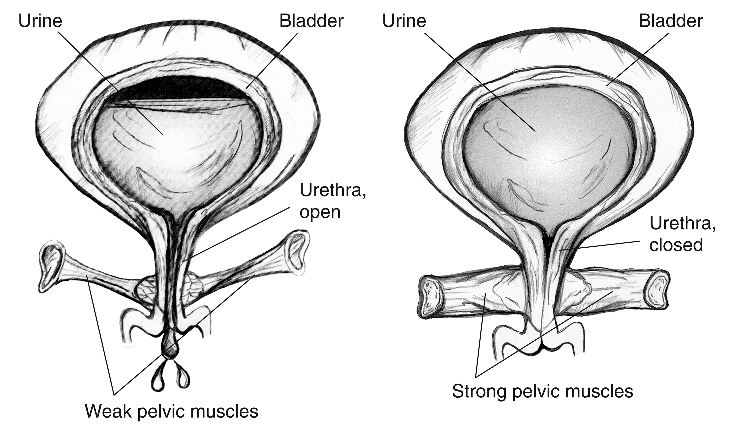
Treat persistent stress leakage after appropriate conservative care.
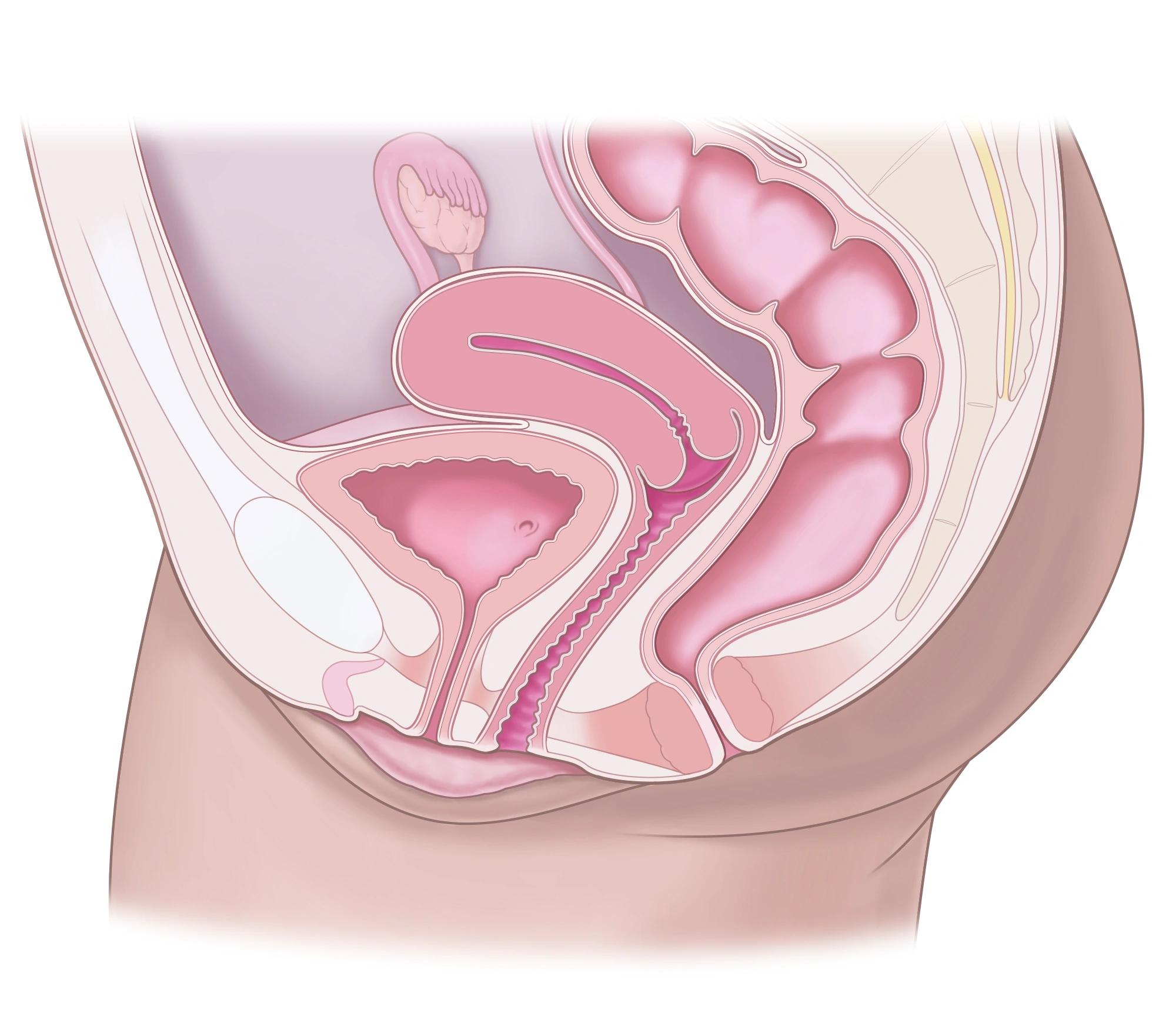
Reading this diagram
The bladder is at the front and connects to the urethra. The uterus and vagina are nearby organs, separate from the urinary tract.
Before
Confirm the leakage mechanism and discuss pregnancy plans, alternatives and implant benefits and risks.
During
Options include a sling, colposuspension or an autologous fascial sling. Techniques and materials differ.
After
Check emptying, pain and leakage; follow activity advice.
What to expect
Reduces stress leakage; it does not necessarily treat urgency.
Risks and limitations
Retention, pain, recurrence or implant complications may require further surgery.
Bladder botulinum toxin injections
Treat selected resistant overactive bladder, with or without neurological disease.
Reading this diagram
The two kidneys are at the top. The ureters lead down to the bladder. Below it, the urethra carries urine out of the body.
Before
Exclude infection and discuss possible self-catheterisation if emptying becomes difficult.
During
Injections are given into the bladder wall during cystoscopy with suitable anaesthesia.
After
Check residual urine and response. Repeat treatment may be needed.
What to expect
May reduce urgency and leakage; the effect is temporary.
Risks and limitations
Urinary infection, blood in urine and retention sometimes requiring catheterisation.
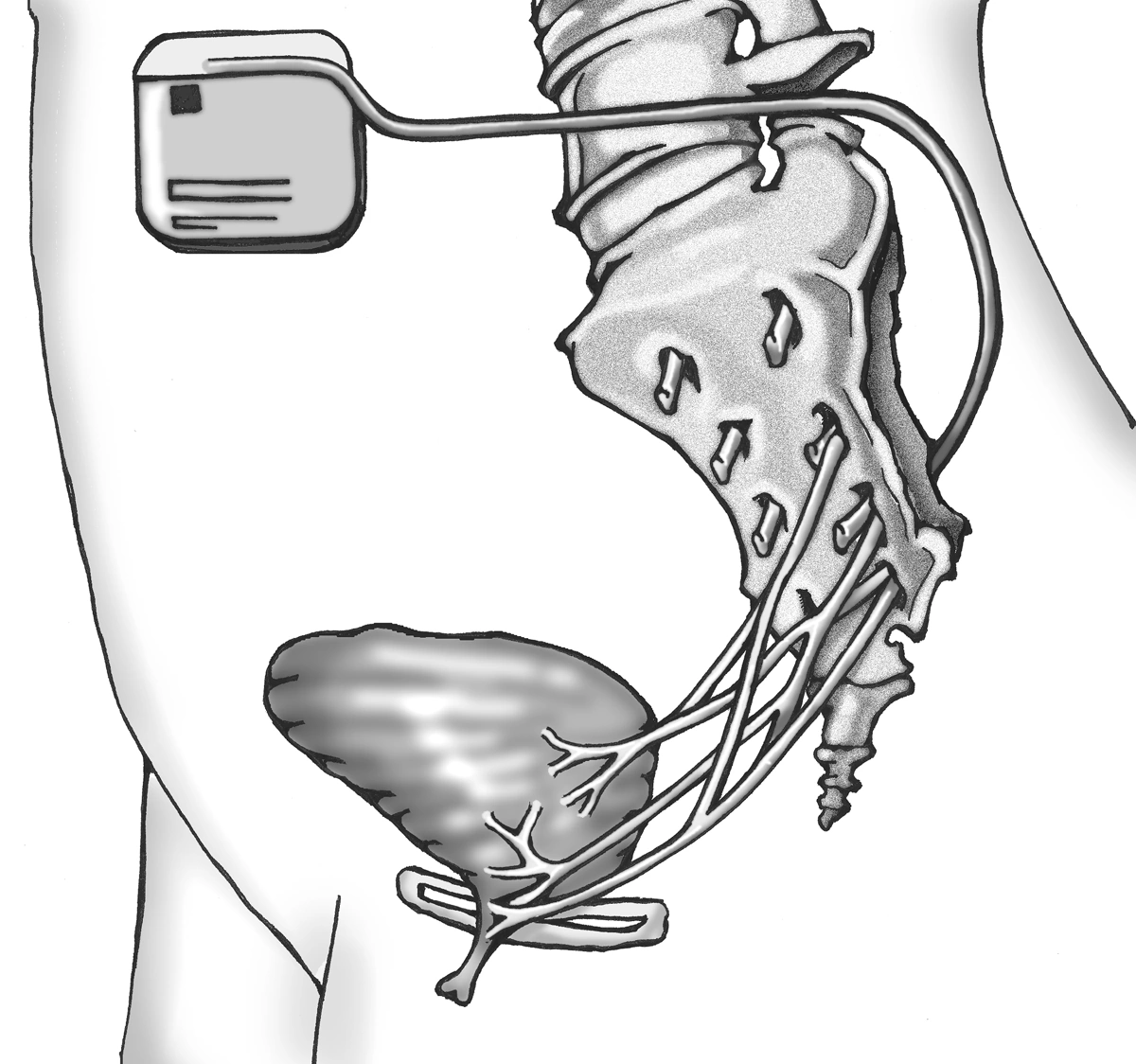
Sacral neuromodulation
A specialist option for selected resistant storage or emptying problems.
Before
Functional assessment and discussion of previous treatment, future imaging and the device.
During
A test phase assesses response before permanent implantation of a stimulator near a sacral nerve.
After
Programming, battery follow-up and implant-site checks.
What to expect
Test improvement helps select patients; benefit can change over time.
Risks and limitations
Pain, infection, displacement or revision may occur; MRI compatibility depends on the device.
Vasectomy
Male contraception intended to be permanent, after informed decision-making.
Reading this diagram
The prostate sits below the bladder. The urethra passes through the prostate and then the penis. The rectum lies behind them.
Before
Discuss alternatives, future parenthood and the uncertain success of any reversal.
During
The vas deferens tubes are interrupted, usually under local anaesthesia.
After
Continue other contraception until laboratory confirmation and medical clearance. Follow the semen-testing protocol.
What to expect
Highly effective after clearance; does not directly reduce testosterone production.
Risks and limitations
Persistent pain, haematoma or rare failure; does not protect against sexually transmitted infections.
Medically indicated circumcision
Treat pathological phimosis or selected recurrent inflammation after assessment.
Reading this diagram
The prostate sits below the bladder. The urethra passes through the prostate and then the penis. The rectum lies behind them.
Before
Confirm indication, alternatives and age-appropriate anaesthesia. A non-retractable foreskin can be normal in children.
During
The foreskin is removed and skin sutured using an individualised approach.
After
Local care, healing review and avoidance of irritating activity until healed as advised.
What to expect
Removes the tight foreskin ring; local sensation can change.
Risks and limitations
Bleeding, infection, healing problems or an unsatisfactory cosmetic result.
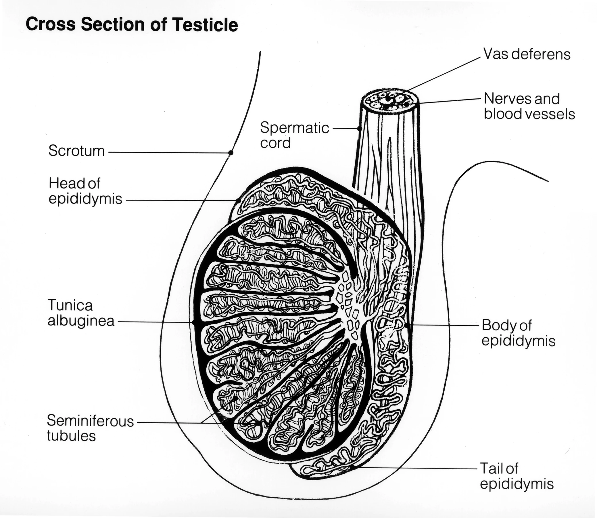
Hydrocele surgery
Treat fluid around the testicle when troublesome or large.
Before
Examination and sometimes ultrasound confirm the cause; children need a specific assessment.
During
The fluid sac is treated surgically through an appropriate approach.
After
Postoperative swelling can occur; monitor the wound and resume effort gradually.
What to expect
Reduces bulk and discomfort; the result is assessed after swelling settles.
Risks and limitations
Haematoma, infection, pain or recurrence may occur.
Varicocele treatment
Consider a clinical varicocele in selected infertility, pain or testicular growth problems.
Before
Examination, semen analysis and couple assessment when seeking pregnancy; not every varicocele requires treatment.
During
Veins are interrupted surgically, often microsurgically, or by embolisation when appropriate.
After
Clinical follow-up; semen changes are assessed over several months.
What to expect
May improve selected parameters or pain without guaranteeing pregnancy.
Risks and limitations
Persistence/recurrence, hydrocele or vascular injury; risks differ by technique.
Orchidopexy — testicular fixation
Bring down an undescended testicle or fix a testicle in torsion; these are different situations.
Before
Undescended testes need early paediatric care. Sudden pain suggesting torsion is an immediate emergency.
During
The testicle is repositioned and fixed according to anatomy and viability; both sides may be fixed in torsion.
After
Review position and size, with follow-up tailored to the original diagnosis.
What to expect
Aims to preserve function; after torsion this depends partly on time to treatment.
Risks and limitations
Atrophy or re-ascent can occur. Surgery for an undescended testicle does not eliminate later cancer risk.
Ask my care team
- Which instructions apply to my medicines and meals?
- Who should I contact after discharge, including outside office hours?
- When can I resume activities and when is follow-up planned?