Clear terms to help you understand your consultation.
20 terms
No term found. Try another spelling.
Active surveillance
Planned monitoring with tests; treatment may be offered if the condition changes.
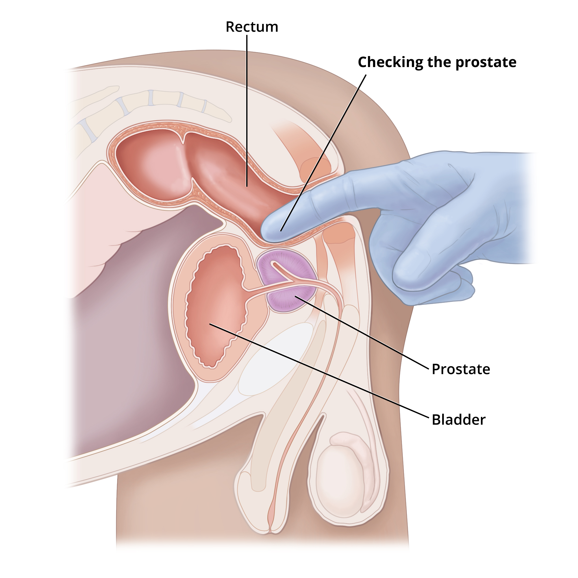
Enlarge illustrationDigital rectal examination of the prostate. Anatomical or procedural reference; it does not by itself depict the condition described.NIDDK / NIHOriginal image labels are in English.National Institute of Diabetes and Digestive and Kidney Diseases, National Institutes of Health.Read the full leaflet
Biopsy
Taking a tissue sample for laboratory examination.
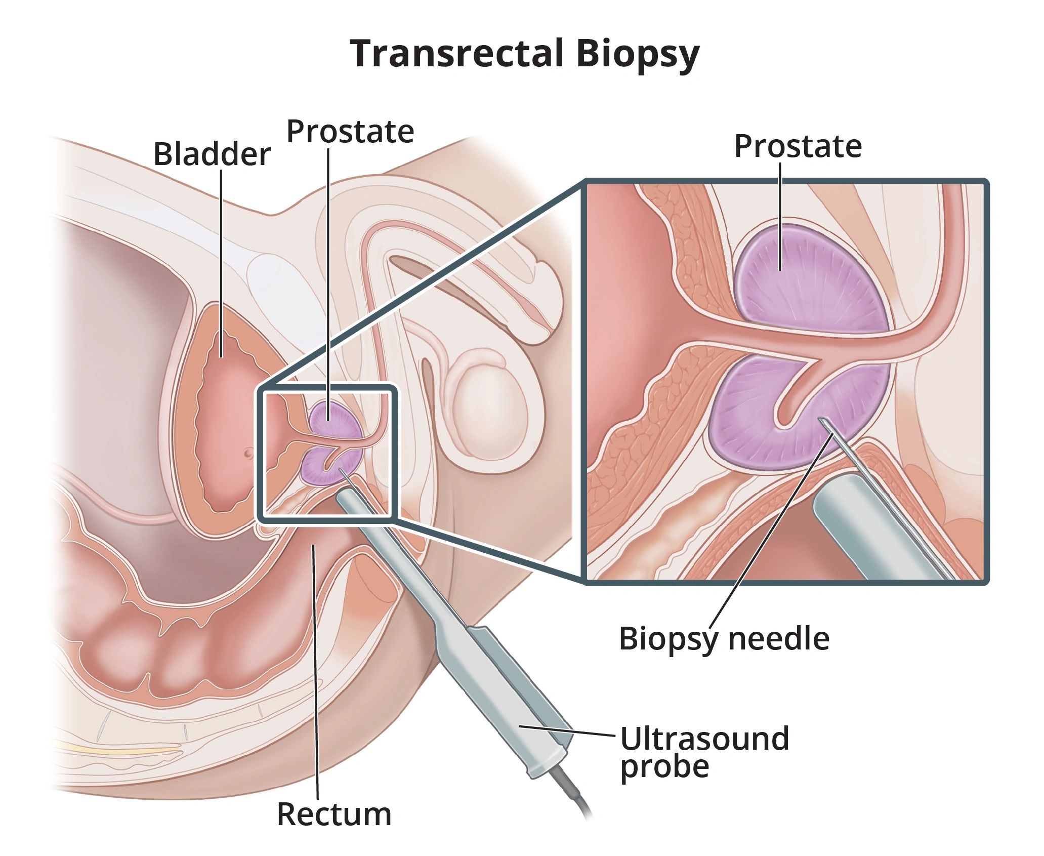
Enlarge illustrationExample of transrectal prostate biopsy. Transperineal biopsy is another approach selected according to context. Anatomical or procedural reference; it does not by itself depict the condition described.NIDDK / NIHOriginal image labels are in English.National Institute of Diabetes and Digestive and Kidney Diseases, National Institutes of Health.Read the full leaflet
Bladder
Organ storing urine before urination.
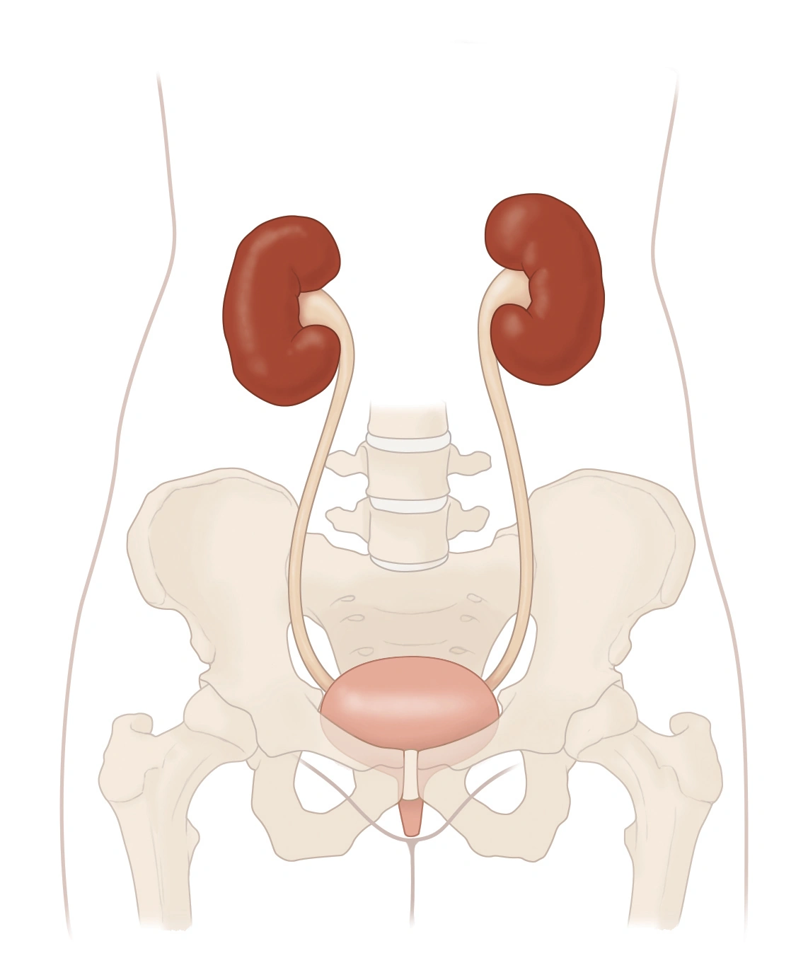
Enlarge illustrationUrinary tract: kidneys, ureters, bladder and urethra. Anatomical or procedural reference; it does not by itself depict the condition described.Reading this diagram
The two kidneys are at the top. The ureters lead down to the bladder. Below it, the urethra carries urine out of the body.
NIDDK / NIHNational Institute of Diabetes and Digestive and Kidney Diseases, National Institutes of Health.Read the full leaflet
Cystoscopy
Viewing the bladder with a camera passed through the urethra.
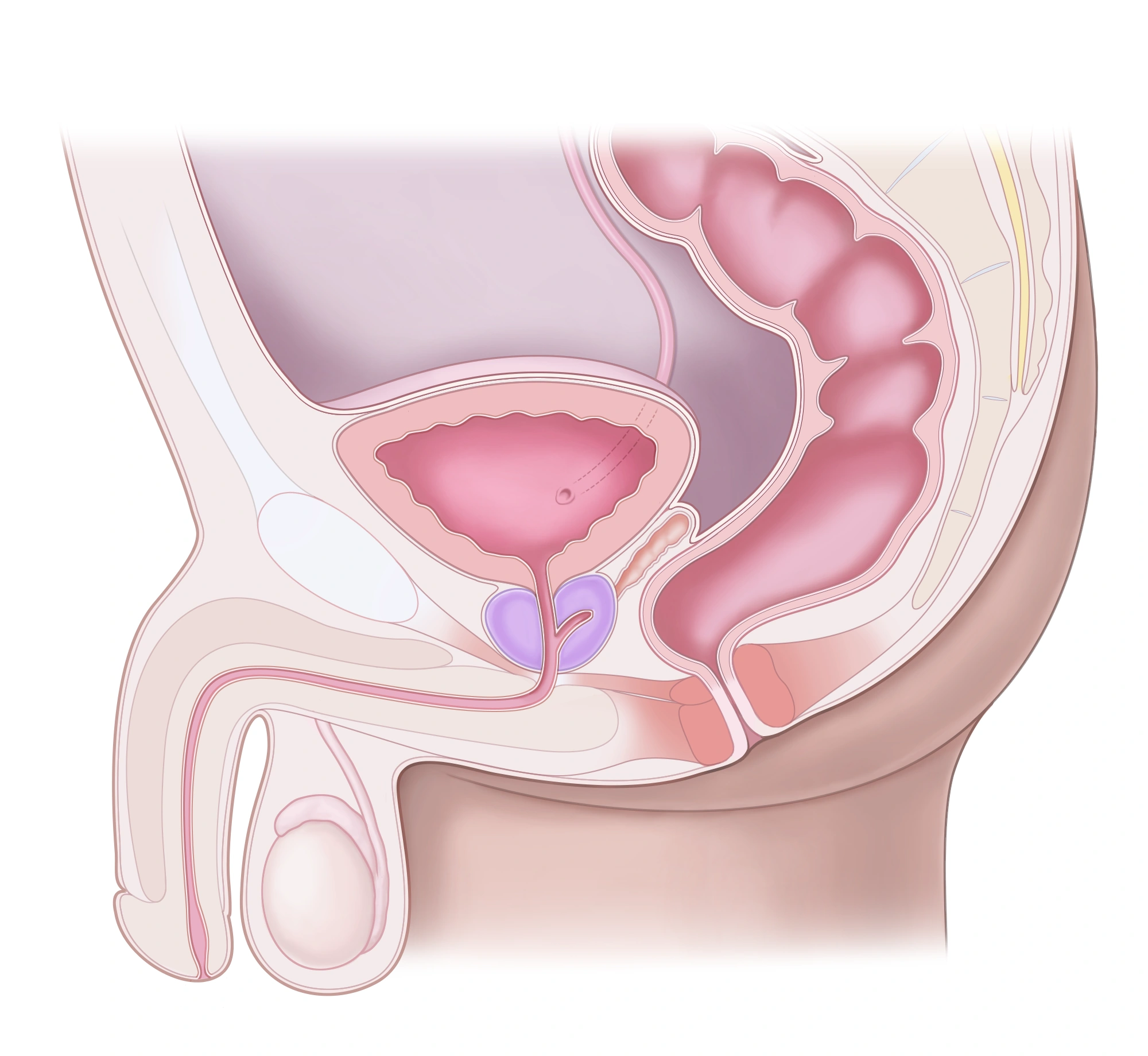
Enlarge illustrationMale pelvic and reproductive anatomy, side view. Anatomical or procedural reference; it does not by itself depict the condition described.Reading this diagram
The prostate sits below the bladder. The urethra passes through the prostate and then the penis. The rectum lies behind them.
NIDDK / NIHNational Institute of Diabetes and Digestive and Kidney Diseases, National Institutes of Health.Read the full leaflet
Haematuria
Blood in urine, visible or detected through testing.
Enlarge illustrationUrinary tract: kidneys, ureters, bladder and urethra. Anatomical or procedural reference; it does not by itself depict the condition described.Reading this diagram
The two kidneys are at the top. The ureters lead down to the bladder. Below it, the urethra carries urine out of the body.
NIDDK / NIHNational Institute of Diabetes and Digestive and Kidney Diseases, National Institutes of Health.Read the full leaflet
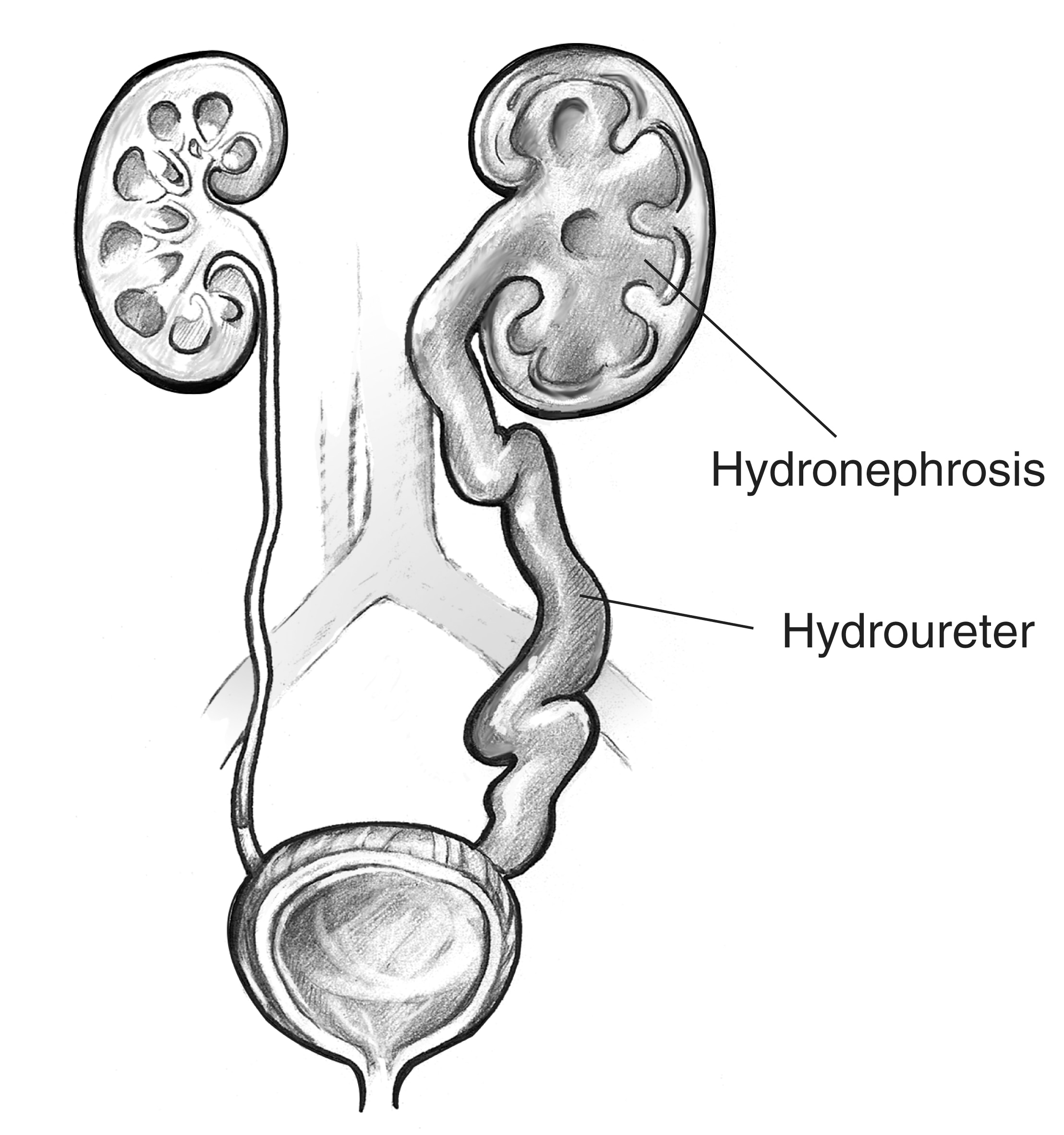
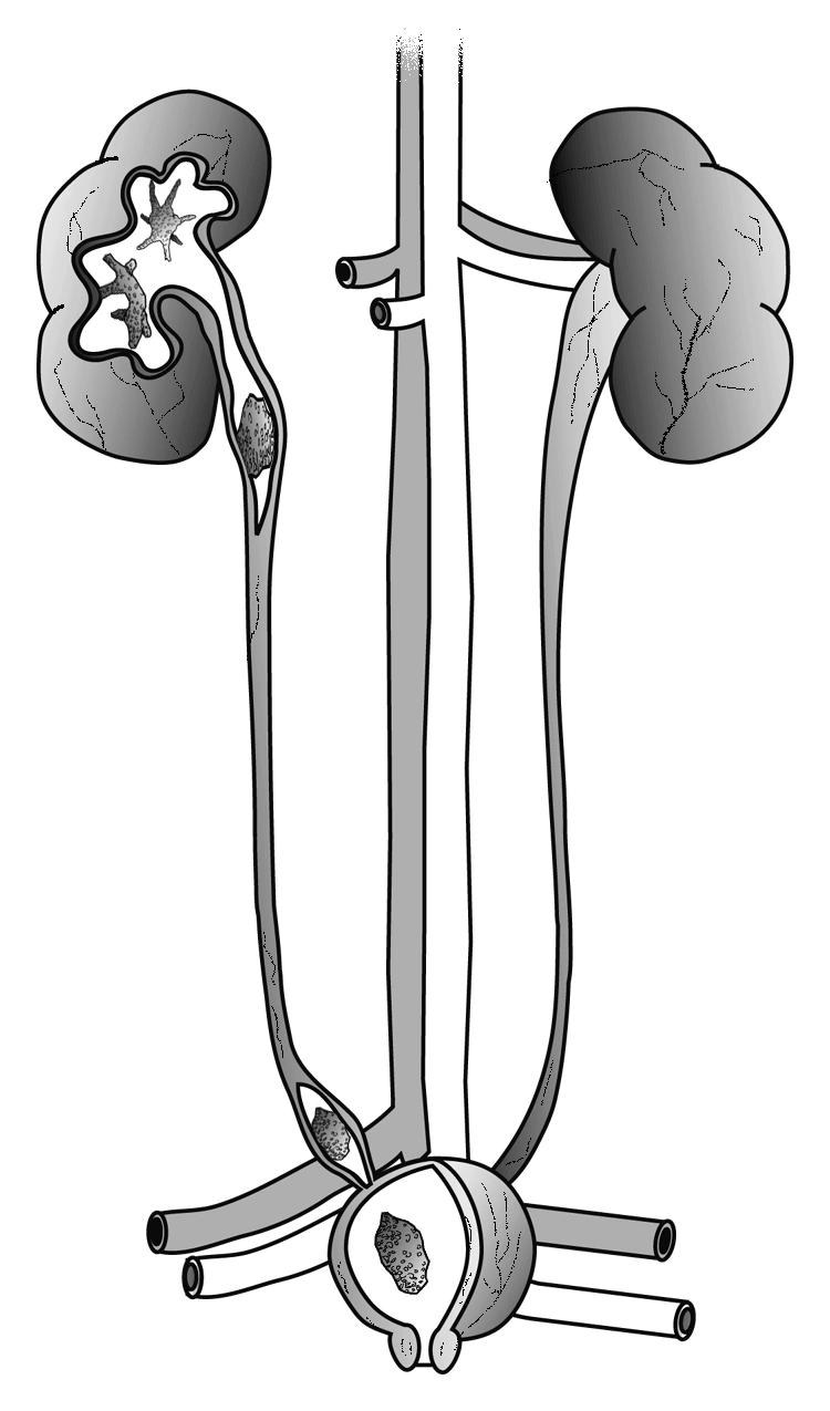
Hydronephrosis
Dilation of the kidney’s urine-collecting spaces.
Enlarge illustrationDilated kidney collecting system and ureter: hydronephrosis and hydroureter. NIDDK / NIHOriginal image labels are in English.National Institute of Diabetes and Digestive and Kidney Diseases, National Institutes of Health.Read the full leaflet
JJ stent
Internal tube between kidney and bladder to help drainage.
Enlarge illustrationUrinary tract: kidneys, ureters, bladder and urethra. Anatomical or procedural reference; it does not by itself depict the condition described.Reading this diagram
The two kidneys are at the top. The ureters lead down to the bladder. Below it, the urethra carries urine out of the body.
NIDDK / NIHNational Institute of Diabetes and Digestive and Kidney Diseases, National Institutes of Health.Read the full leaflet
Kidney
Organ that filters blood and produces urine.
Enlarge illustrationKidney and nephron: blood filtration and urine formation. Anatomical or procedural reference; it does not by itself depict the condition described.NIDDK / NIHOriginal image labels are in English.National Institute of Diabetes and Digestive and Kidney Diseases, National Institutes of Health.Read the full leaflet
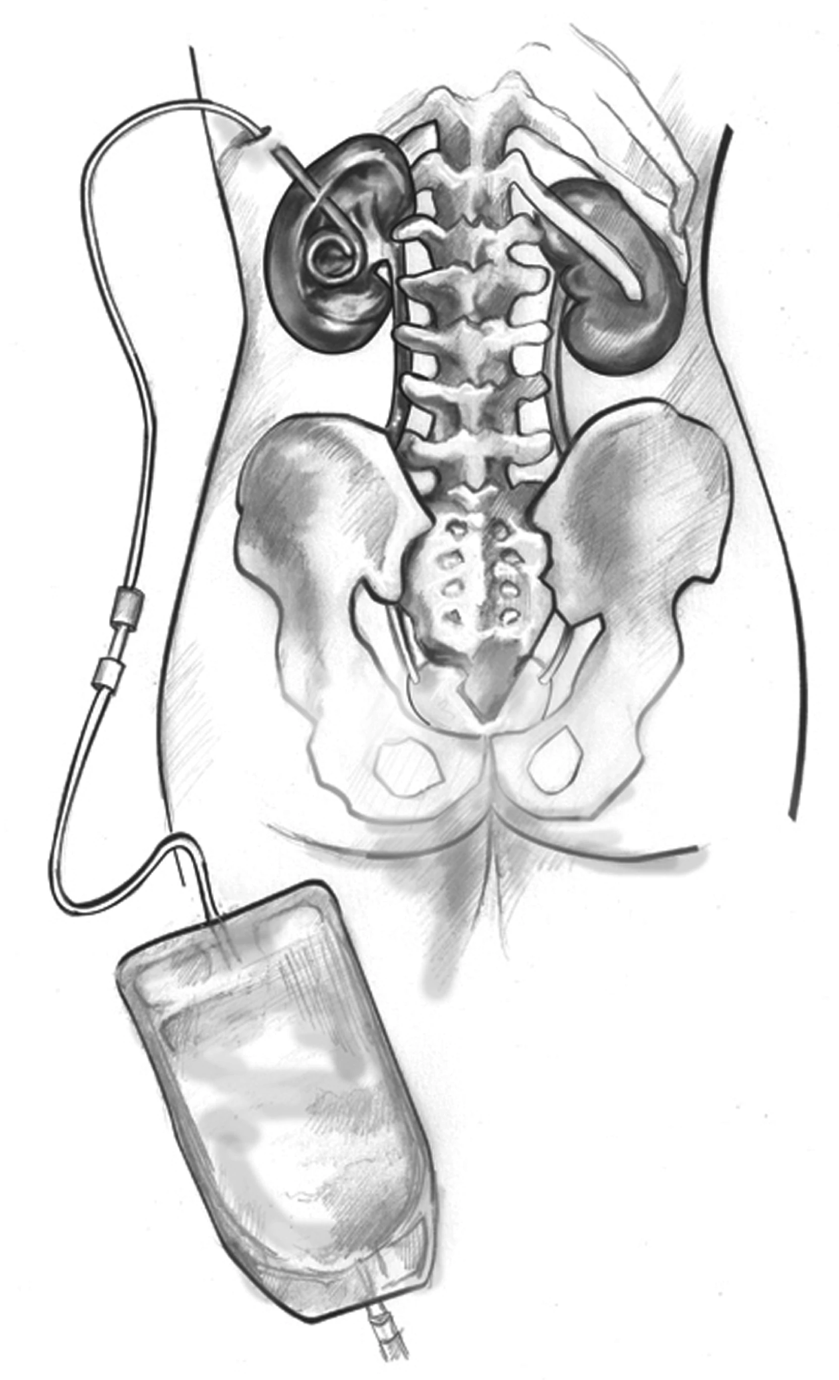
Nephrostomy
Tube draining urine from the kidney through the skin.
Enlarge illustrationNephrostomy tube connecting the kidney to an external collection bag. NIDDK / NIHNational Institute of Diabetes and Digestive and Kidney Diseases, National Institutes of Health.Read the full leaflet
Post-void residual
Urine left in the bladder after urination.
Enlarge illustrationUrinary tract: kidneys, ureters, bladder and urethra. Anatomical or procedural reference; it does not by itself depict the condition described.Reading this diagram
The two kidneys are at the top. The ureters lead down to the bladder. Below it, the urethra carries urine out of the body.
NIDDK / NIHNational Institute of Diabetes and Digestive and Kidney Diseases, National Institutes of Health.Read the full leaflet
Prostate
Gland below the bladder surrounding the first part of the urethra.
Enlarge illustrationMale pelvic and reproductive anatomy, side view. Anatomical or procedural reference; it does not by itself depict the condition described.Reading this diagram
The prostate sits below the bladder. The urethra passes through the prostate and then the penis. The rectum lies behind them.
NIDDK / NIHNational Institute of Diabetes and Digestive and Kidney Diseases, National Institutes of Health.Read the full leaflet
PSA
Prostate protein measured in blood. One result alone does not diagnose cancer.
Enlarge illustrationDigital rectal examination of the prostate. Anatomical or procedural reference; it does not by itself depict the condition described.NIDDK / NIHOriginal image labels are in English.National Institute of Diabetes and Digestive and Kidney Diseases, National Institutes of Health.Read the full leaflet
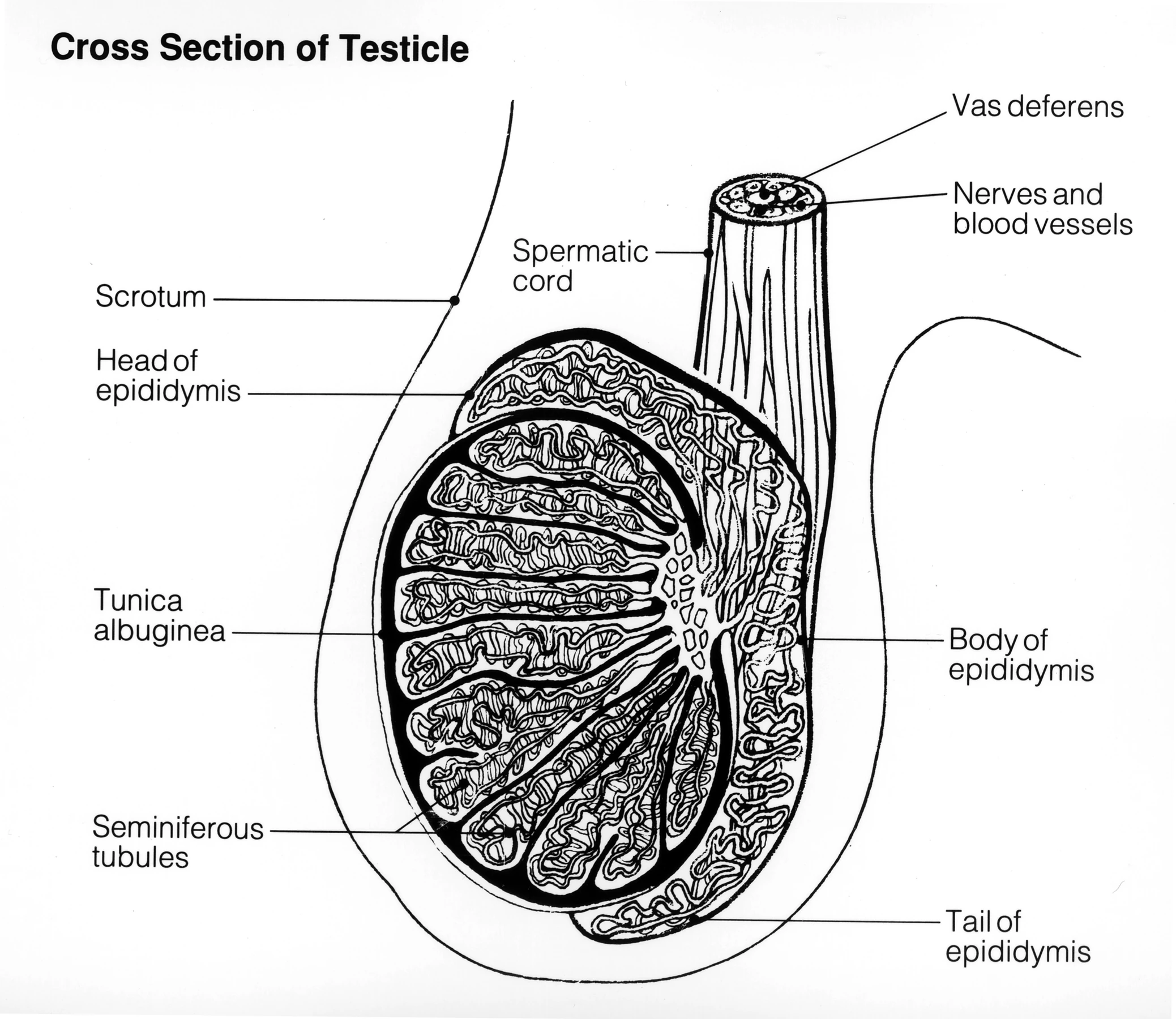
Semen analysis
Examination of semen as part of fertility assessment.
Enlarge illustrationCross-section of the testis and epididymis: anatomical landmarks. Anatomical or procedural reference; it does not by itself depict the condition described.NCI / NIHOriginal image labels are in English.National Cancer InstituteRead the full leaflet
Ureter
Tube carrying urine from the kidney to the bladder.
Enlarge illustrationUrinary tract: kidneys, ureters, bladder and urethra. Anatomical or procedural reference; it does not by itself depict the condition described.Reading this diagram
The two kidneys are at the top. The ureters lead down to the bladder. Below it, the urethra carries urine out of the body.
NIDDK / NIHNational Institute of Diabetes and Digestive and Kidney Diseases, National Institutes of Health.Read the full leaflet
Urethra
Tube through which urine leaves the bladder.
Enlarge illustrationUrinary tract: kidneys, ureters, bladder and urethra. Anatomical or procedural reference; it does not by itself depict the condition described.Reading this diagram
The two kidneys are at the top. The ureters lead down to the bladder. Below it, the urethra carries urine out of the body.
NIDDK / NIHNational Institute of Diabetes and Digestive and Kidney Diseases, National Institutes of Health.Read the full leaflet
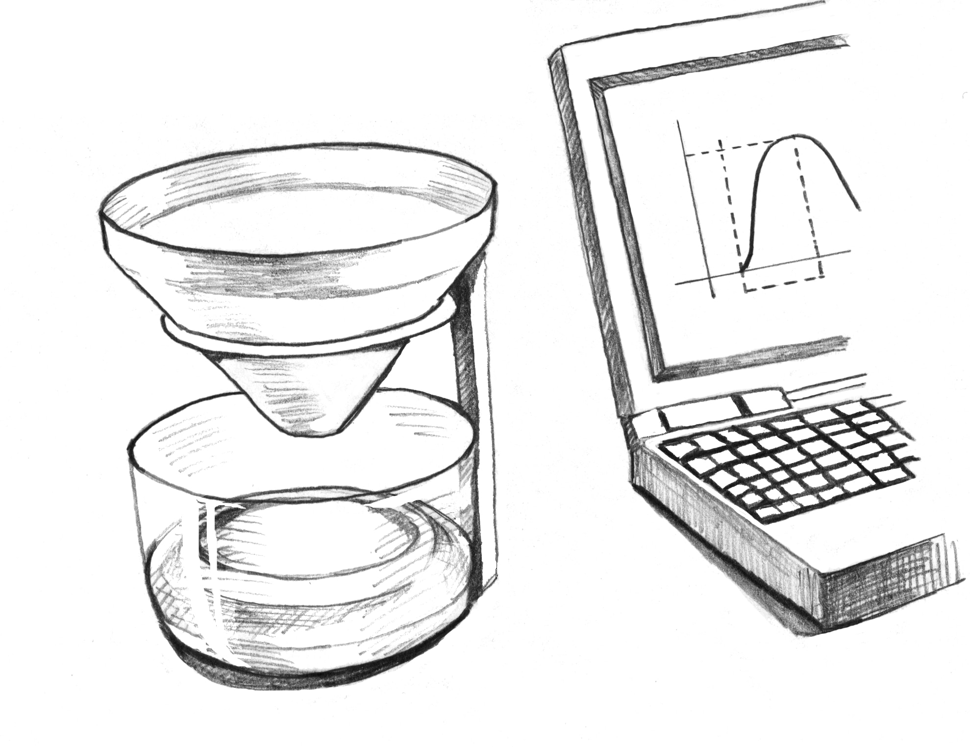
Enlarge illustrationUroflowmeter: urine collection and flow recording. The illustrated curve is not a diagnostic result. NIDDK / NIHNational Institute of Diabetes and Digestive and Kidney Diseases, National Institutes of Health.Read the full leaflet
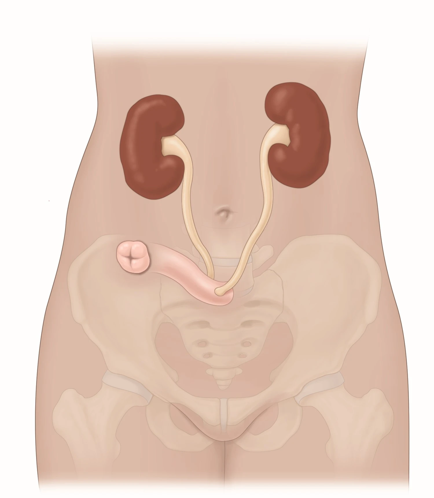
Urostomy
Surgically created opening allowing urine to leave the body.
Enlarge illustrationCross-section of the testis and epididymis: anatomical landmarks. Anatomical or procedural reference; it does not by itself depict the condition described.NCI / NIHOriginal image labels are in English.National Cancer InstituteRead the full leaflet