
Dr Ali Beddouche · Tests at the practice
Prepare for my test
Choose your scheduled test to find preparation, steps and documents.
Your individual practice instructions take priority. Do not change your medicines yourself.
Urinary tract ultrasound
Read the leaflet and keep its PDF
Your test, step by step
Before the test
Follow the practice’s instructions about bladder filling. Mention any fluid restriction and avoid holding urine until it hurts. Bring previous reports.
During
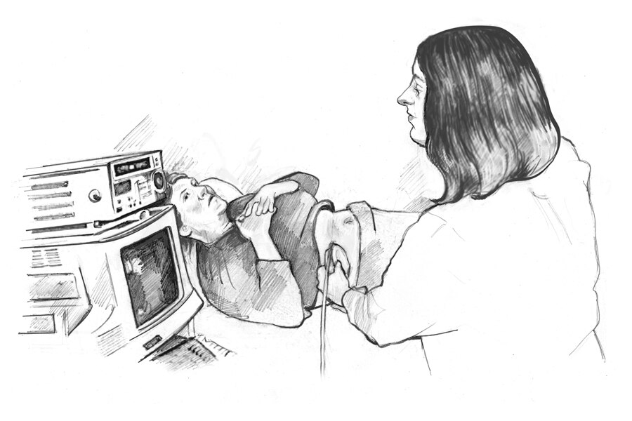
A probe with gel moves over the abdomen and sides. A scan after urinating may measure how much urine remains in the bladder.
Afterwards and results
Ask for the report and how the findings relate to your symptoms. A normal ultrasound does not replace any other tests that may be needed.
Guy’s and St Thomas’ NHS · Kidney and bladder ultrasound
Choose another testUrine flow test
Read the leaflet and keep its PDF
Your test, step by step
Before the test
Arrive with a normal urge to urinate, without overfilling your bladder. Tell the team if holding urine is difficult or you have a fluid restriction.
During
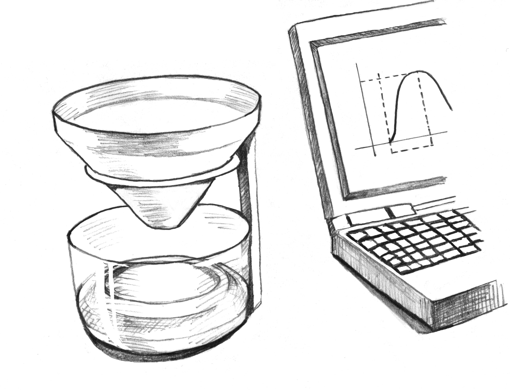
Urinate in private into the device, in your usual position when possible. Do not strain or deliberately interrupt the stream.
Afterwards and results
An ultrasound may measure urine left in the bladder. The curve is interpreted with your symptoms; the test may be repeated if it did not reflect your usual urination.
Princess Alexandra Hospital · Uroflow study
Choose another testFlexible cystoscopy
Read the leaflet and keep its PDF
Reading this diagram
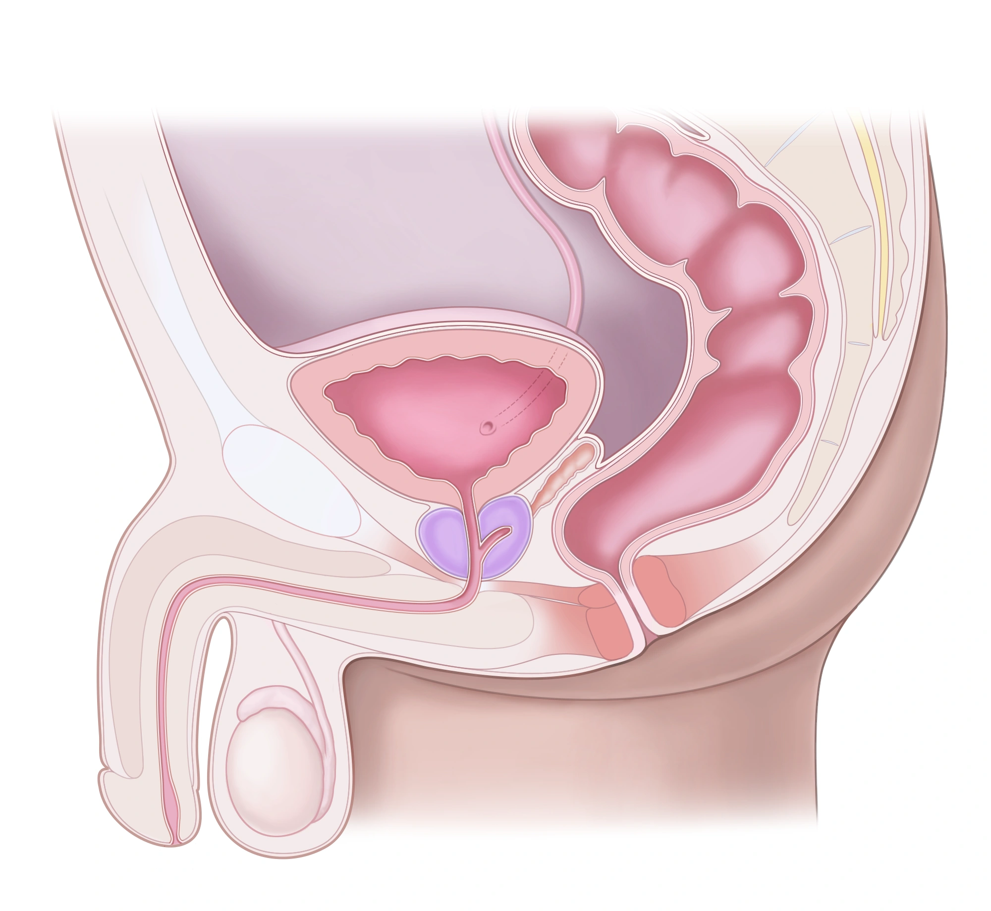
The prostate sits below the bladder. The urethra passes through the prostate and then the penis. The rectum lies behind them.
Your test, step by step
Before the test
Fasting is usually unnecessary with local anaesthetic alone unless you are instructed otherwise. Report fever, urinary burning, allergies and medicines. Complete any requested urine test; do not change medicines yourself.
During
After applying the gel, the doctor gently passes the camera through the urethra. Filling the bladder may make you feel the urge to urinate. Report pain or significant discomfort.
Afterwards and results
Mild burning or small traces of blood may occur briefly. Ask about the findings and follow-up. Fever, chills, worsening pain, heavy bleeding or inability to urinate require prompt medical advice.
My recovery, care and follow-up
NHS · Preparing for cystoscopy · Gloucestershire Hospitals · Flexible cystoscopy
Choose another testTransrectal prostate biopsy
Read the leaflet and keep its PDF
Your test, step by step
Before the test
Review your prescription and preparation instructions. Confirm the antibiotic, its timing and the plan for your usual medicines. Report infection or allergies and arrange someone to accompany you home.
During
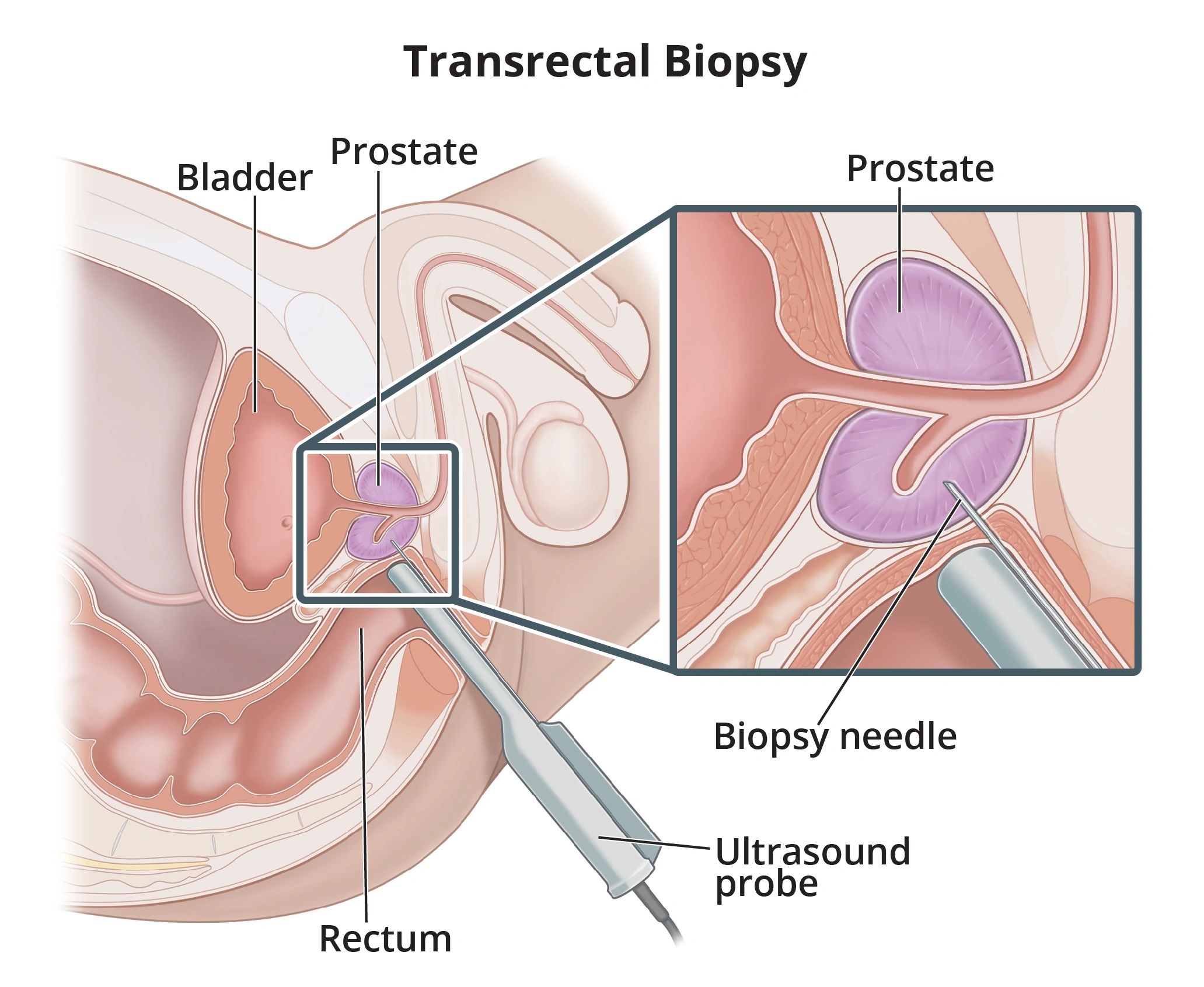
The ultrasound probe is placed in the rectum. After local anaesthesia, the needle crosses the rectal wall to collect prostate tissue samples for laboratory analysis.
Afterwards and results
Keep your discharge instructions and arrange the results appointment. Fever, chills, feeling unwell, inability to urinate or significant bleeding require urgent assessment. See the dedicated leaflet for detailed guidance.
My recovery, care and follow-up
AFU / Urofrance · Biopsie échoguidée de prostate · EAU · Prostate cancer, diagnostic evaluation
Choose another testKeep with you
- Your appointment details and instructions.
- Your medicines and allergy list.
- Your available test results, images and reports.